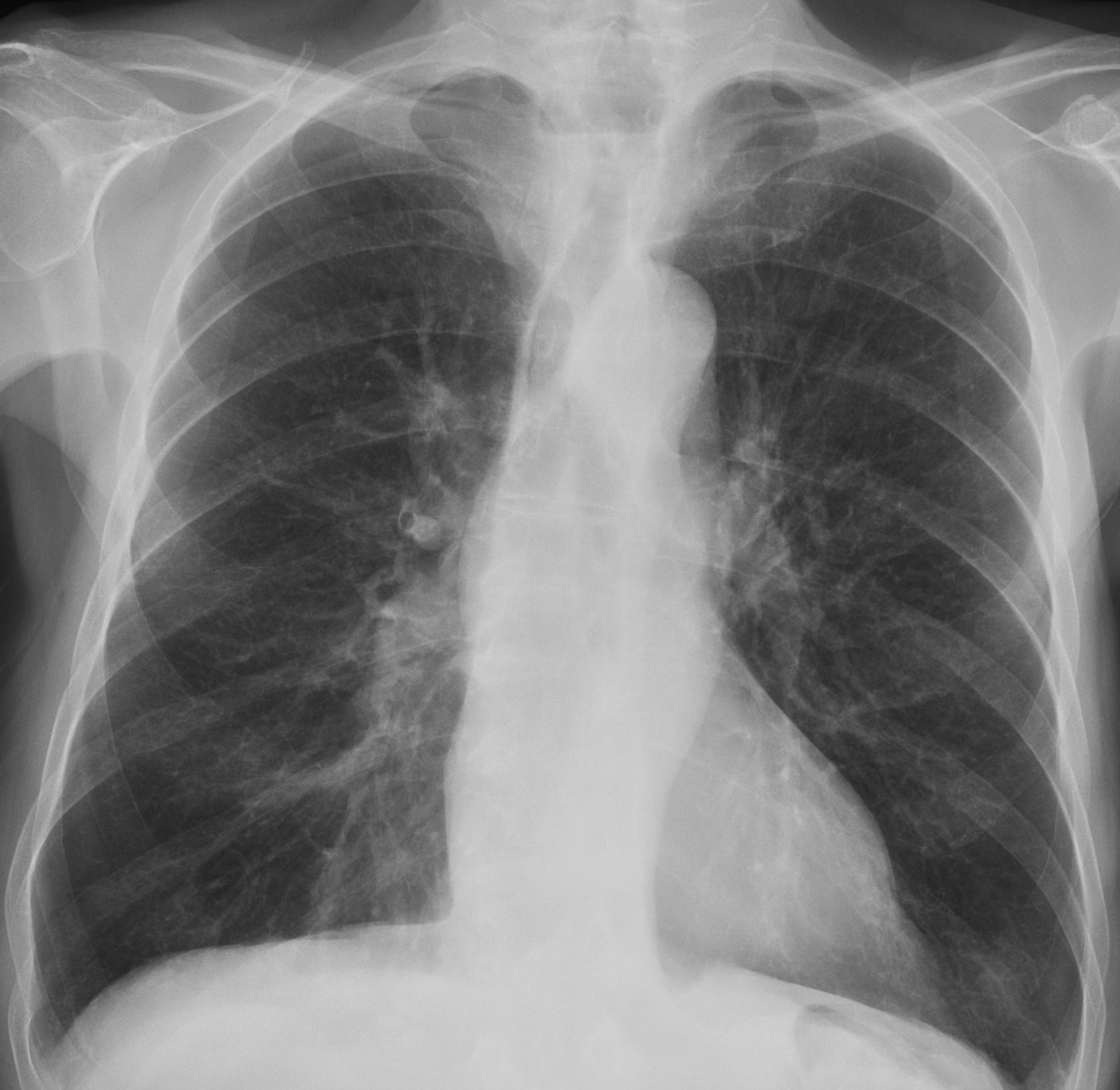
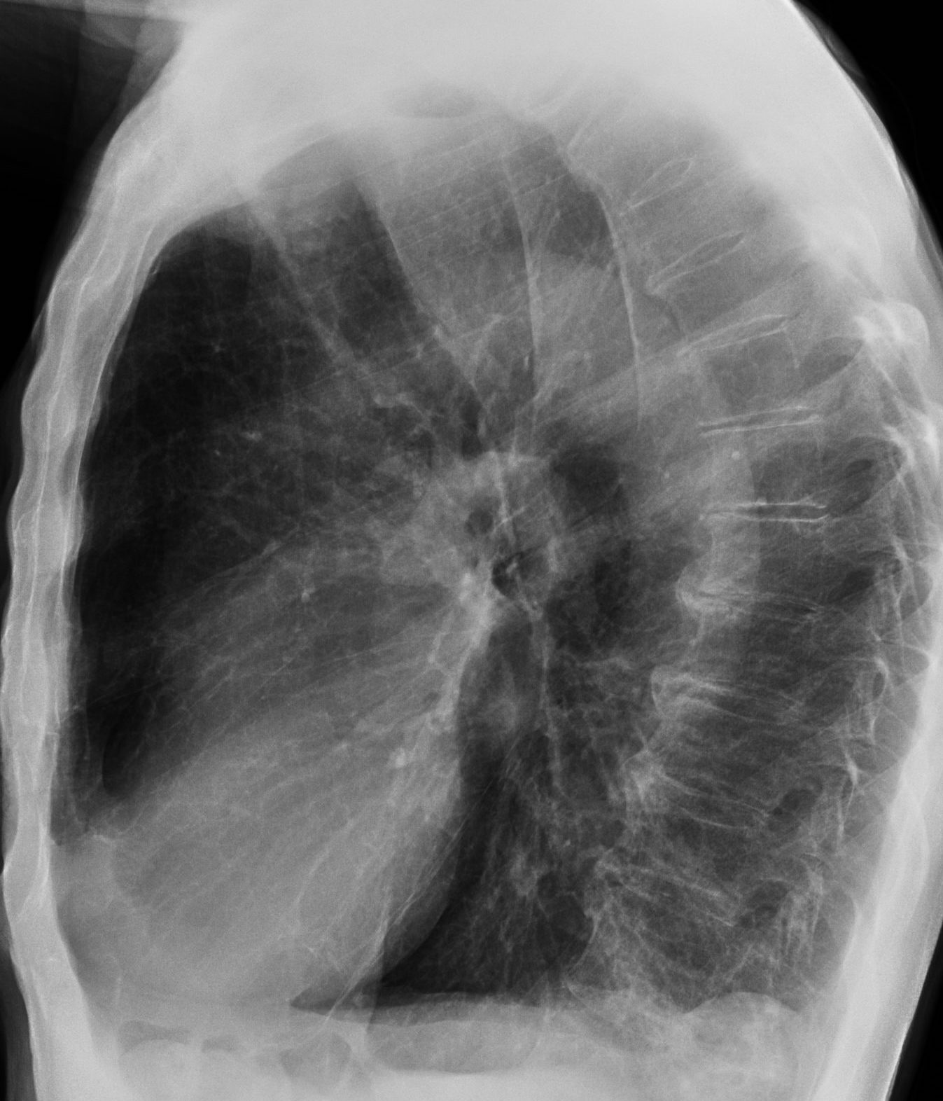
Today I present images of an 89-year-old man with COPD and occasional pulmonary infections. What do you see?
Check the images below, leave your thoughts in the comments section and come back on Friday for the answer.
Findings: chest radiographs show signs of COPD, with in increased anterior clear space and flattening of the diaphragm. A small basal infiltrate is seen in the lateral view (B, arrow). The main finding is a mediastinal air-fluid level visible in both projections (A-B, red arrows).
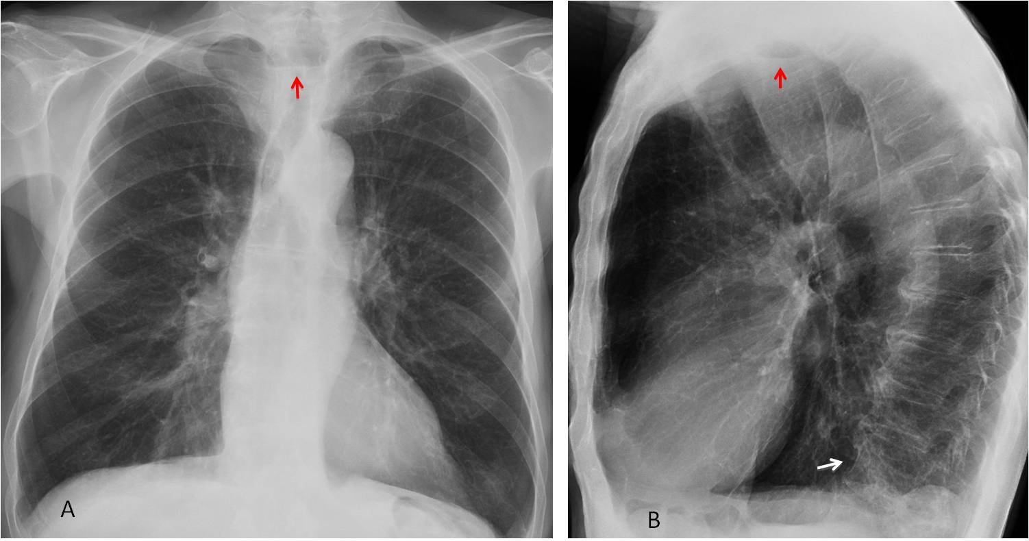
The air-fluid level is more evident in the cone down views (C-D, red arrows) and it occupies Raider triangle in the lateral view.
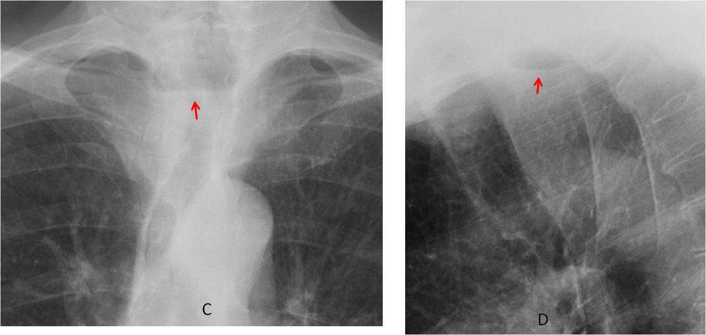
The appearance suggests either oesophageal obstruction or Zenker diverticulum, although I favour the second possibility, given the absence of dysphagia, the rounded appearance of the lesion and the long-standing symptoms.
Oesophagogram (not available) showed a Zenker diverticulum, confirmed at surgery.
Final diagnosis: Zenker diverticulum
Congratulation to MK, who was the first to suggest the diagnosis.
Teaching point: This is a nice case of occupation of Raider triangle by esophageal pathology (the other one being congenital abnormalities of aortic arch)