Since you will be tired after the ECR Congress, I am showing an easy case. These images were taken during a routine check-up of a healthy 55-year-old woman. What do you think?
Check the images below, leave your thoughts in the comments section and come back on Friday for the answer.
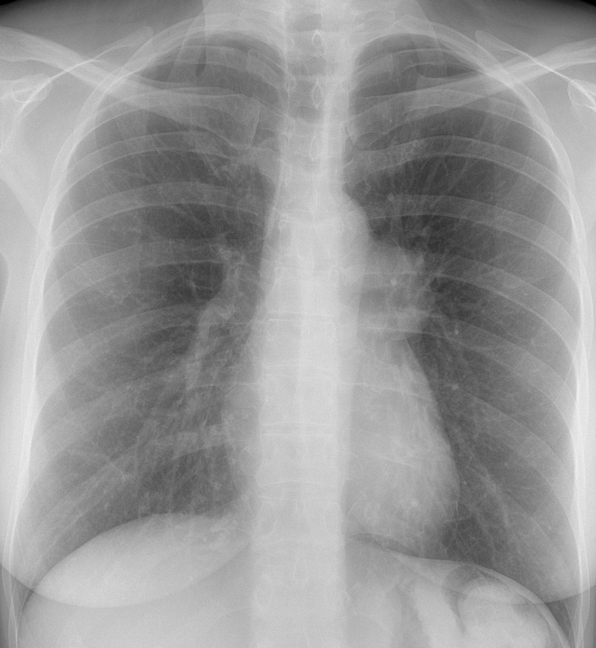
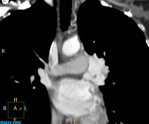
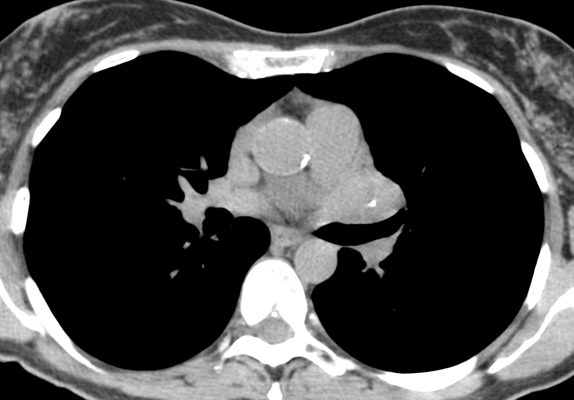
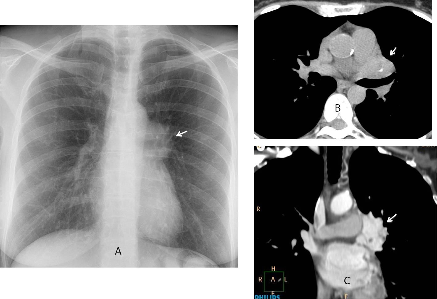
Findings: the PA chest radiograph shows an enlarged right hilum (A, arrow). Axial unenhanced CT confirms the hilar mass (B, arrow) with a central calcification. Enhanced coronal CT shows marked contrast enhancement of the mass (C, arrow).
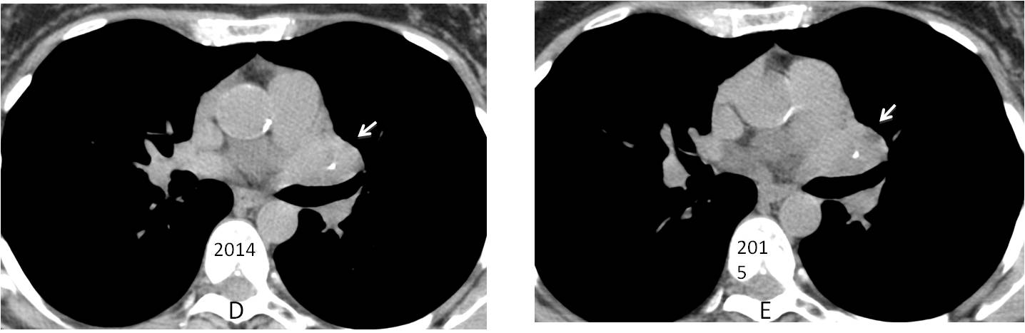
This case has an interesting history. The patient had a routine check-up in 2014 with an unenhanced CT (D) diagnosed as normal. Repeated CT in 2015 (E) was reported as unchanged. Fortunately, a chest radiograph was taken at that time and the hilar abnormality was discovered. It prompted an enhanced CT, which clearly showed the markedly enhancing hilar mass.
The main differential diagnosis of markedly enhancing masses in the chest is limited: Castleman’s disease, carcinoid, glomus tumour and thyroid. Because the location of the mass, I made the diagnosis of Castleman’s.
The patient was operated on and a benign carcinoid was found.
Final diagnosis: central carcinoid tumour of the lung.
Congratulations to all of you who participated and offered a correct differential diagnosis: tr, MK, 19Medicus83 and ren.
Teaching point: this case emphasises one basic principle: “always think of a rare manifestation of a common disease than a common manifestation of a rare disease”. I chose to ignore it and made the wrong diagnosis.