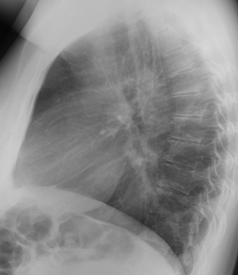
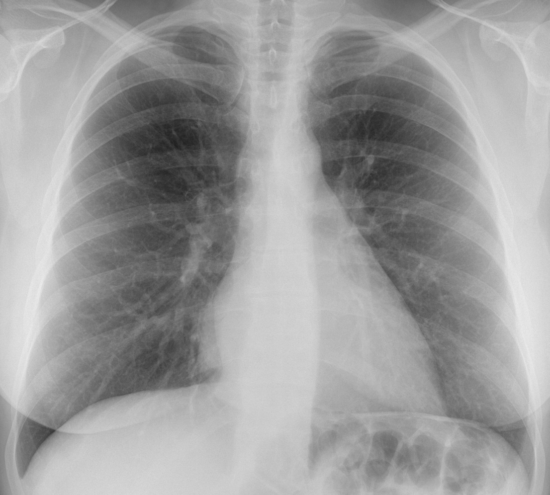
Spring is here and it makes us want to present easy cases. Today we are showing preoperative radiographs for ankle trauma in a 47-year-old woman.
What do you see?
Check the images below, leave your thoughts in the comments section and come back on Friday for the answer.
Findings: the PA radiograph shows a well-defined line (A, arrow) paralleling the right heart border, suggesting a mediastinal mass. The lateral view shows occupation of the retrocardiac space (B, arrows), locating the mass in the lower middle mediastinum.
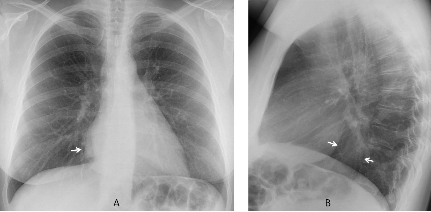
The first diagnostic option should be an airless hiatus hernia, followed by oesophageal lesions such as duplication cyst, wall tumour or varices.
Coronal and axial enhanced CT shows a solid mass adherent to the oesophageal wall (C-D, arrows). Endoscopy confirmed an oesophageal parietal tumour with mild ulceration.
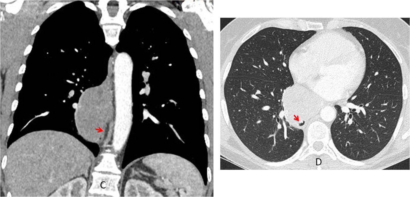
Final diagnosis: oesophageal parietal tumour (GIST)
Congratulations to all of you who placed the mass in the lower middle mediastinum, led by maurizio franz, who was the first.
Teaching point: remember that any abnormality should be properly evaluated and placed. Once this is done, the differential diagnosis is easy. This is a simple case and some of you made it complicated. Don’t forget the KISS method!