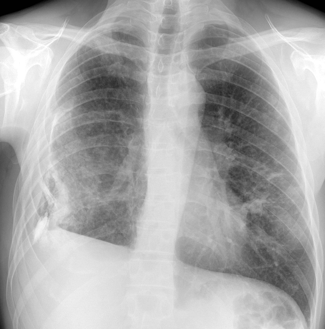
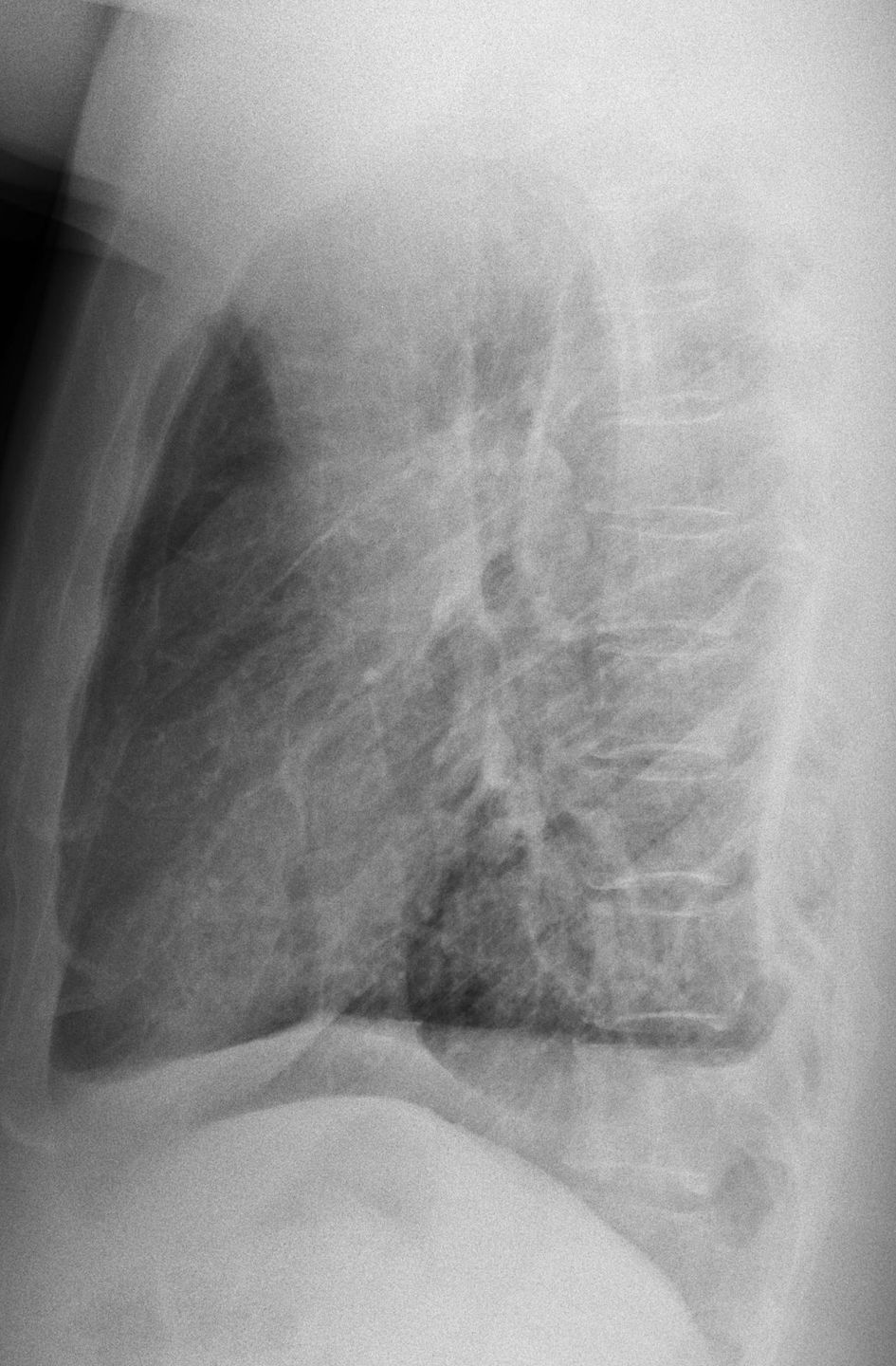
Today I am showing chest radiographs of a 47-year-old man with fever and moderate dyspnoea. What do you see?
Check the images below, leave your thoughts in the comments section and come back on Friday for the answer.
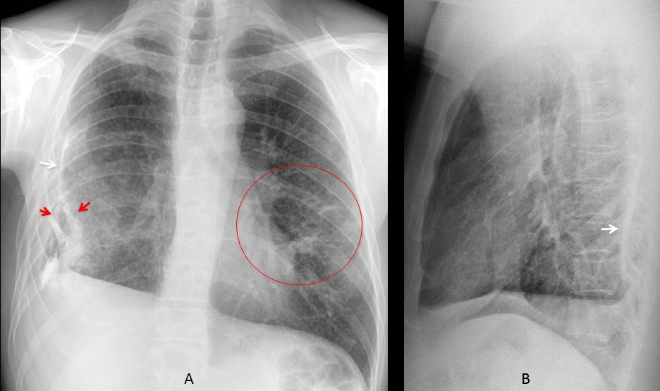
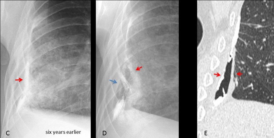
Findings: Chest radiographs show a small right hemithorax secondary to chronic calcified pleural thickening (A-B, arrows). The lower aspect of the pleura is split (A, red arrows), with air between both pleural layers. The appearance is pathognomonic of a bronchopleural fistula. There are pulmonary infiltrates in the left lung (A, circle).
Cone down PA view taken six years earlier shows only calcified pleural thickening (D, arrow). Compare with the present radiograph (E) and the coronal CT (F).
Unenhanced axial CT shows the global appearance of the pleura and B-P fistula (G-E, arrows) as well as the infiltrates in the left lung. BK was found in the bronchial lavage.
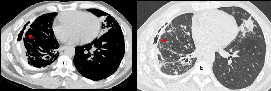
Final diagnosis: Bronchopleural fistula secondary to chronic pleural TB.
Congratulations to MK, who was the first to mention the correct diagnosis
Teaching point: air in the pleural space may come from the outside or the inside. If no invasive procedure has been performed, the most likely diagnosis is B-P fistula, excluding the rare occurrence of pleural infection by gas-forming organisms.