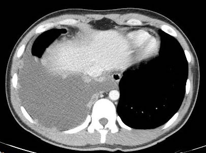
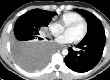
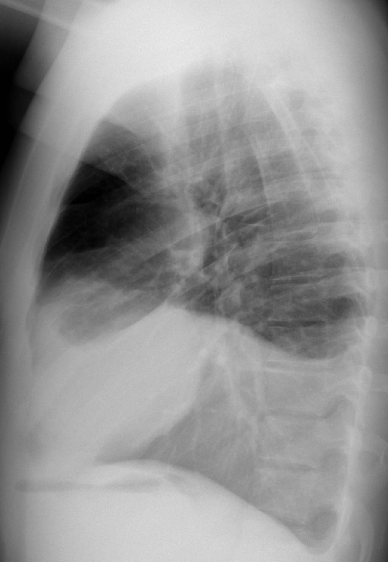
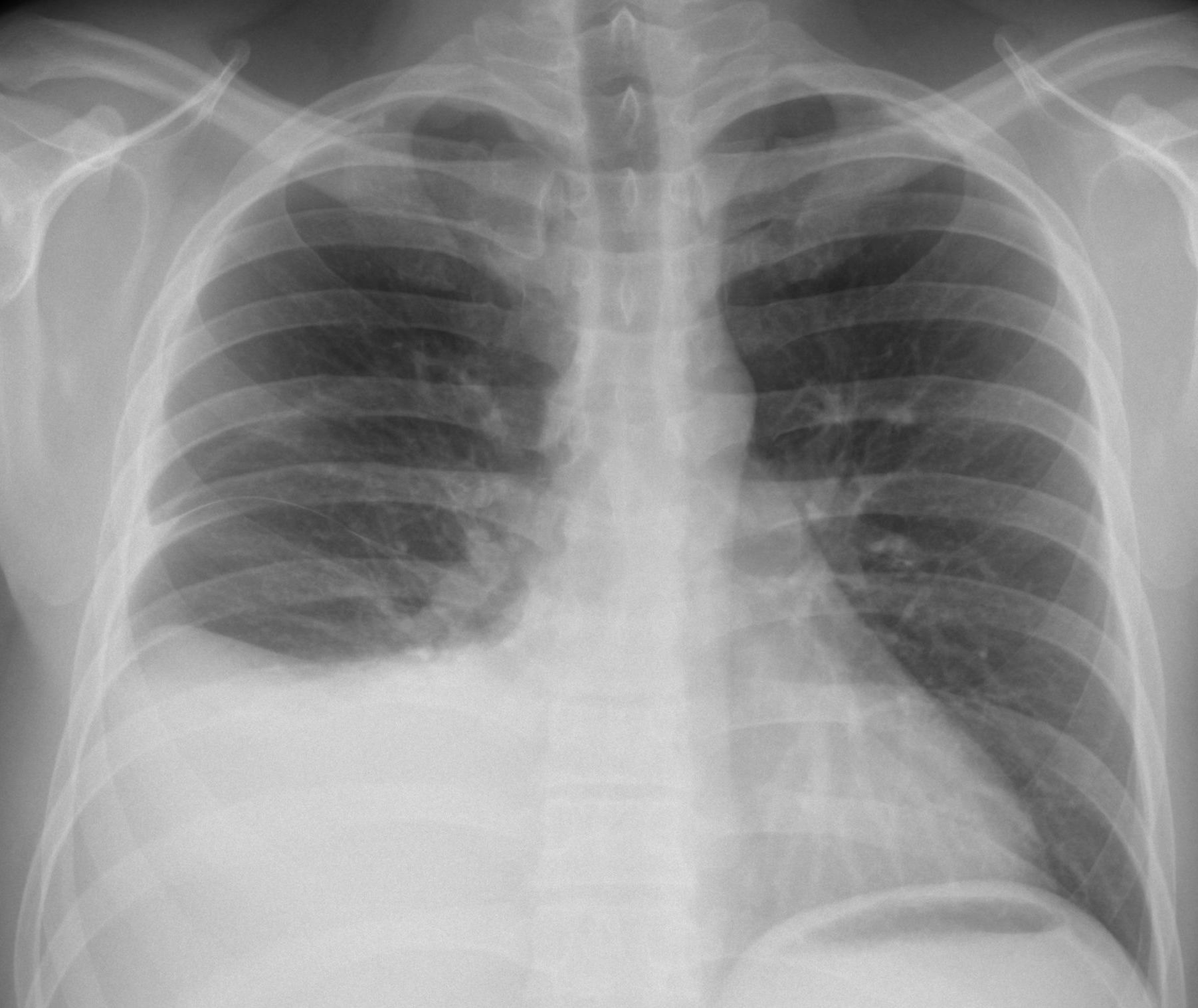
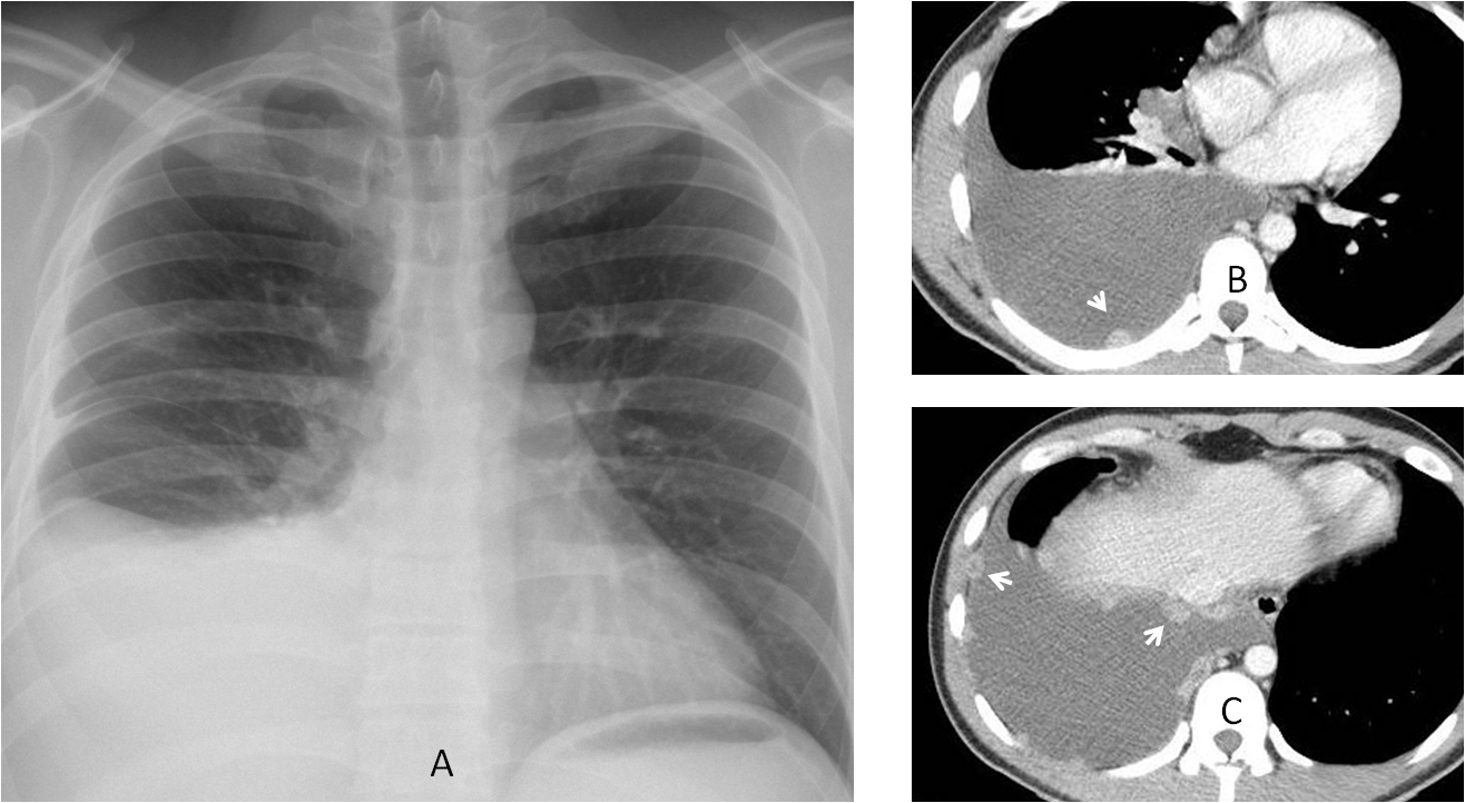
Today we are presenting radiographs and CT images of a 35-year-old male tourist from Venezuela, who came to the ER with pain in the right hemithorax for the last five days. No fever.
As usual, check the images below, leave your thoughts in the comments section and come back on Friday for the answer.
1. Mesothelioma
2. TB
3. Metastases
4. Any of the above
Findings: the chest radiograph (A) shows a right pleural effusion. Axial CT shows numerous enhancing pleural nodules (B-C, arrows).
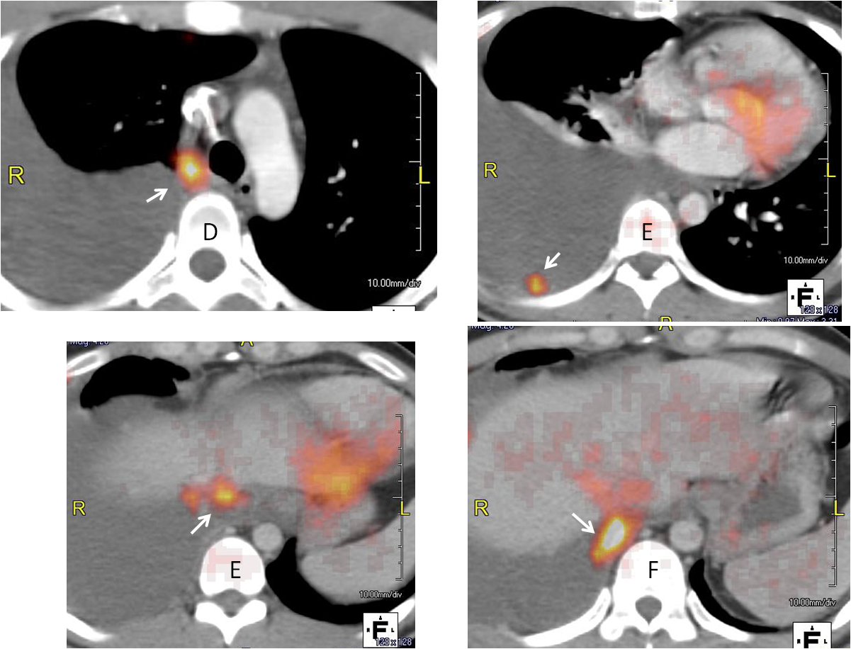
PET-CT shows uptake of the nodules (D-G, arrows)
When I saw this case, I thought of malignancy as the first possibility (either metastases or lymphoma). To my surprise and shame, pleural biopsy revealed casseifying granulomas, BK positive. I did not consider TB because I had never seen pleural nodules in TB and there was no lung involvement.
I wanted to share this case with you because it is a first for me, although this presentation of pleural TB has been described. I am not surprised about the positive PET-CT, because I have seen it many times in active TB.
Final diagnosis: tuberculous pleuritis.
Congratulations to Genchi Bari, who made the diagnosis in unequivocal terms.
Teaching point: remember that TB is a great imitator. It should be included in the differential diagnosis of malignancy of the chest.