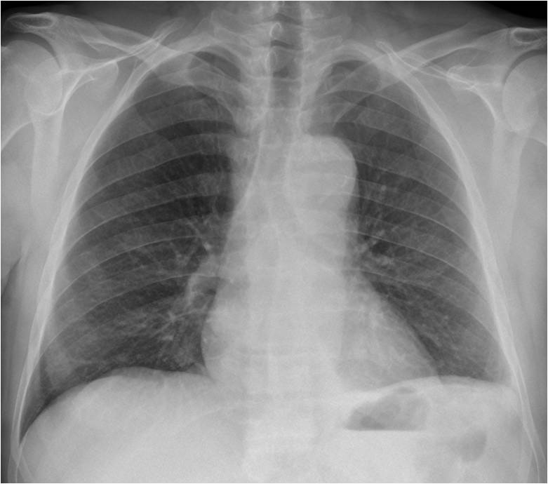
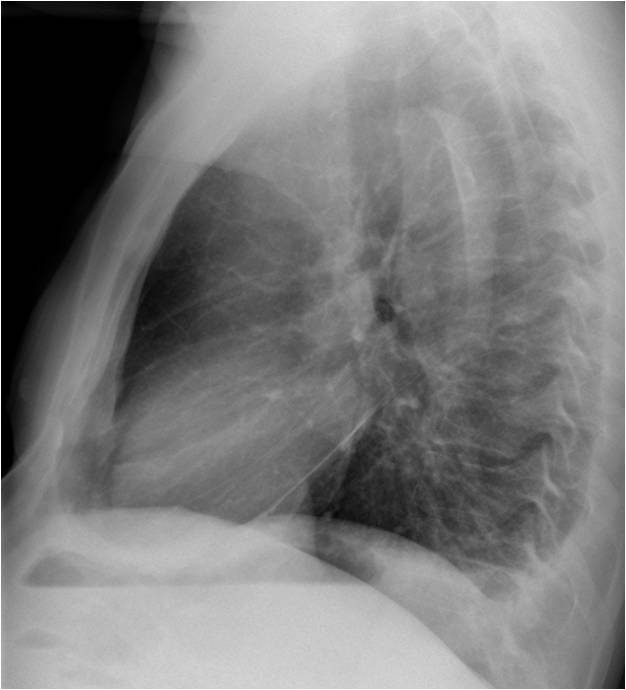
Today I am showing a case from my good friend Jordi Andreu. Radiographs belong to a 52-year-old man with chest pain.
What do you see?
Check the images below, leave your thoughts in the comments section, and come back on Friday for the answer.
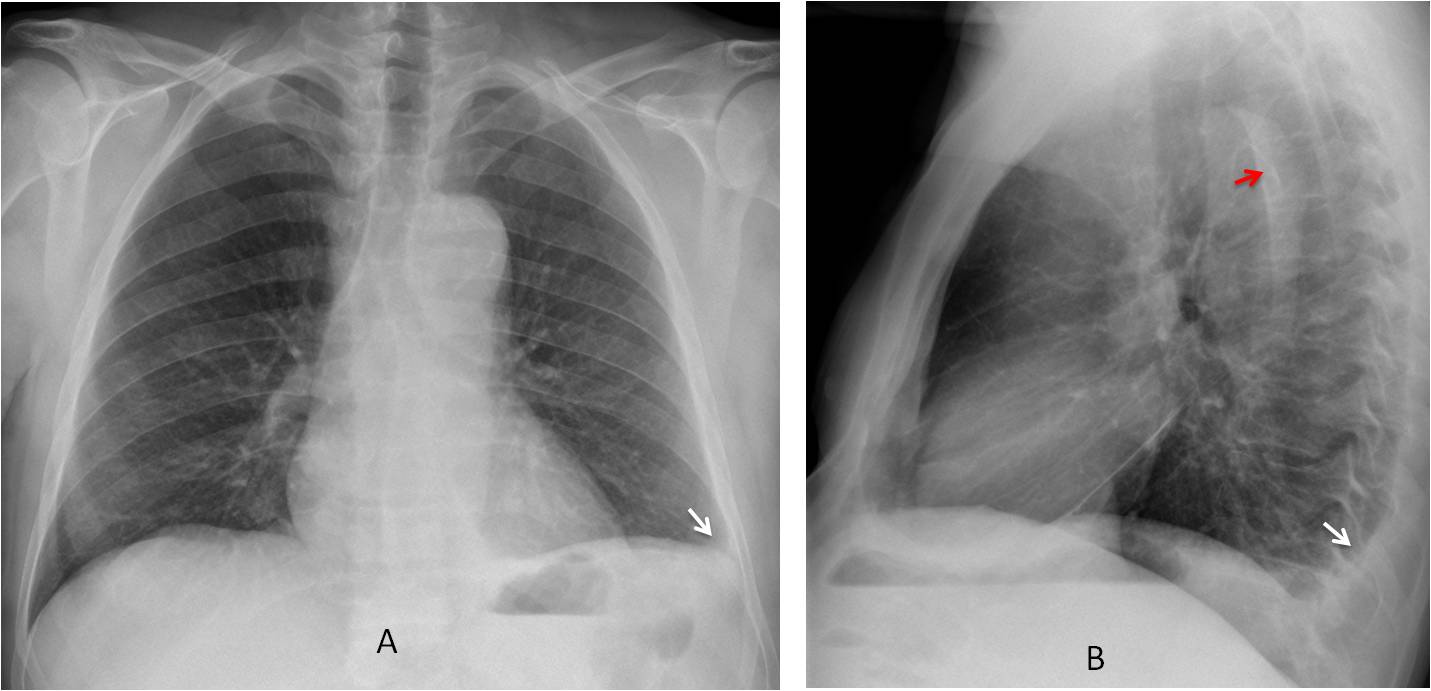
Findings: PA and lateral radiographs show a small left pleural effusion (A-B, arrows). Curvilinear intimal calcium within the aortic lumen is seen in the lateral view (B, red arrow). This finding is highly suspicious of aortic dissection.
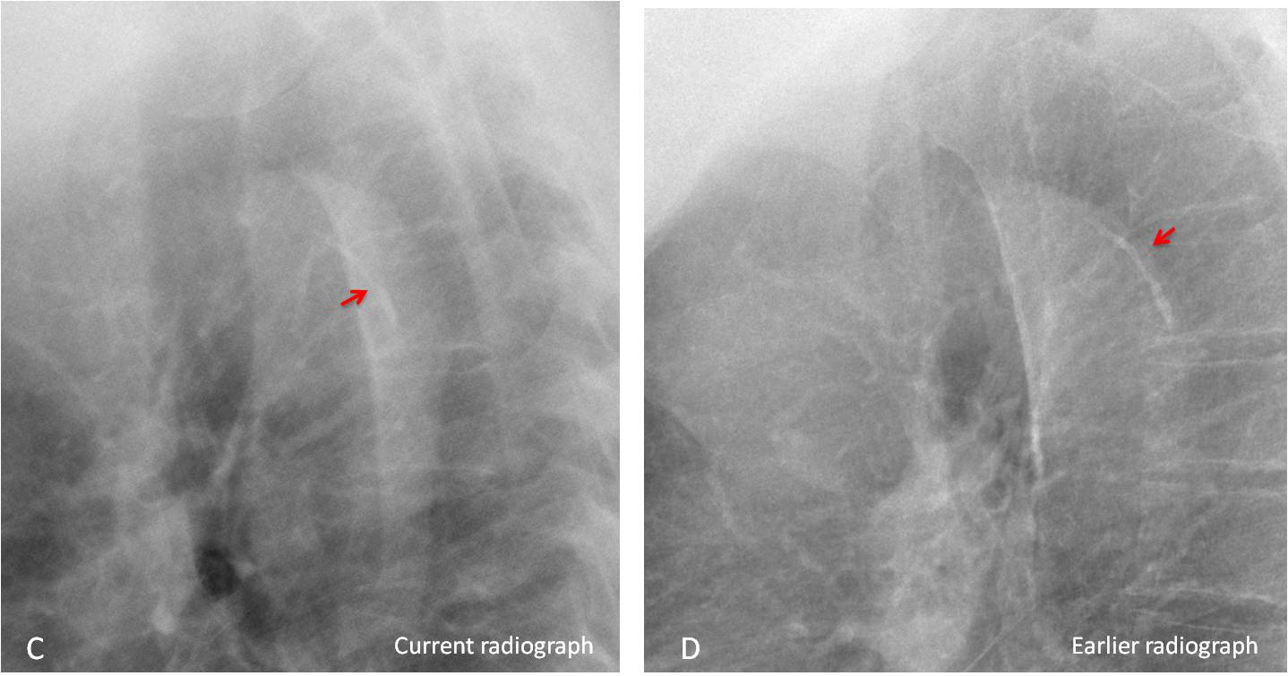
Comparison with a previous lateral view confirms the displacement of the intimal calcium (C, red arrow), which was located in the periphery in the earlier film (D, red arrow).
A previous axial CT shows the calcium in the aortic wall (E, arrow). Current CT shows the dissection surrounding the aortic lumen with the calcium displaced inward (F, arrow). Enhanced CT confirms the findings (G, arrows).
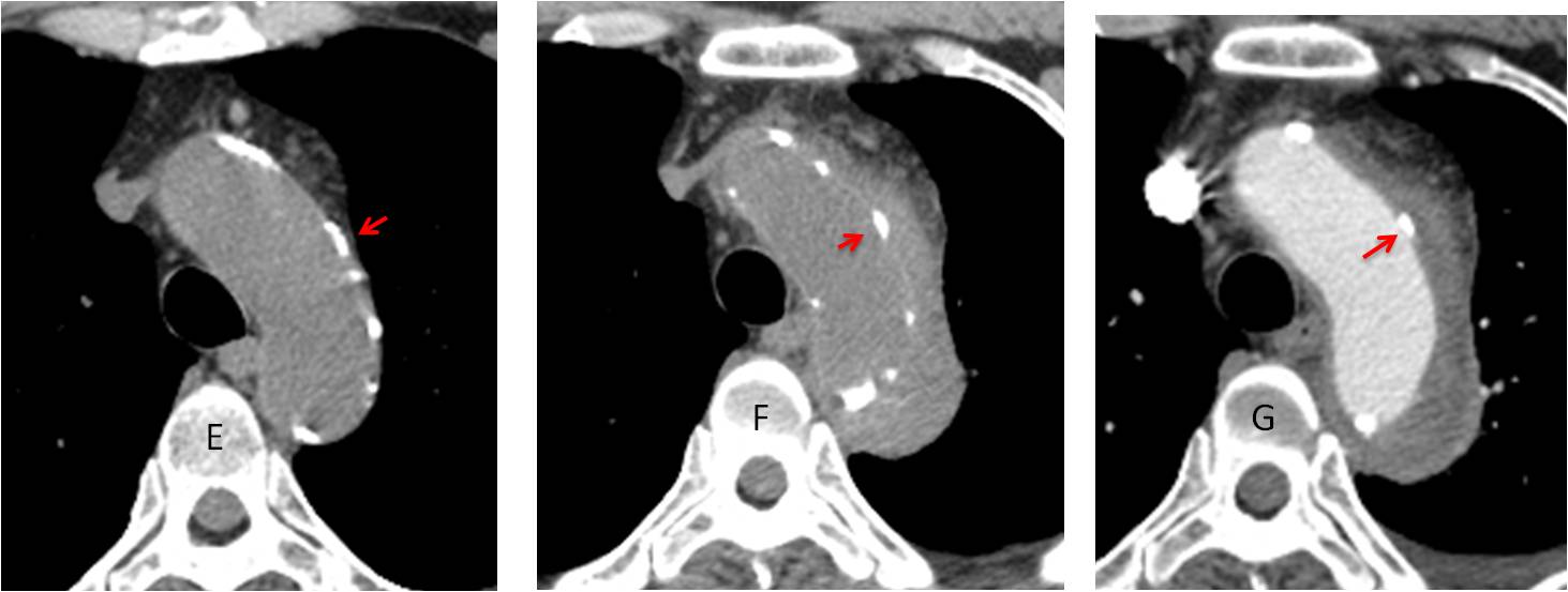
Final diagnosis: acute type B aortic dissection.
Congratulations to all of you who made the diagnosis, led by MK, who was to first to mention it.
Teaching point: this is a nice example of the value of the lateral view and of comparing with previous studies. This led to suggest the right diagnosis, later confirmed by CT.