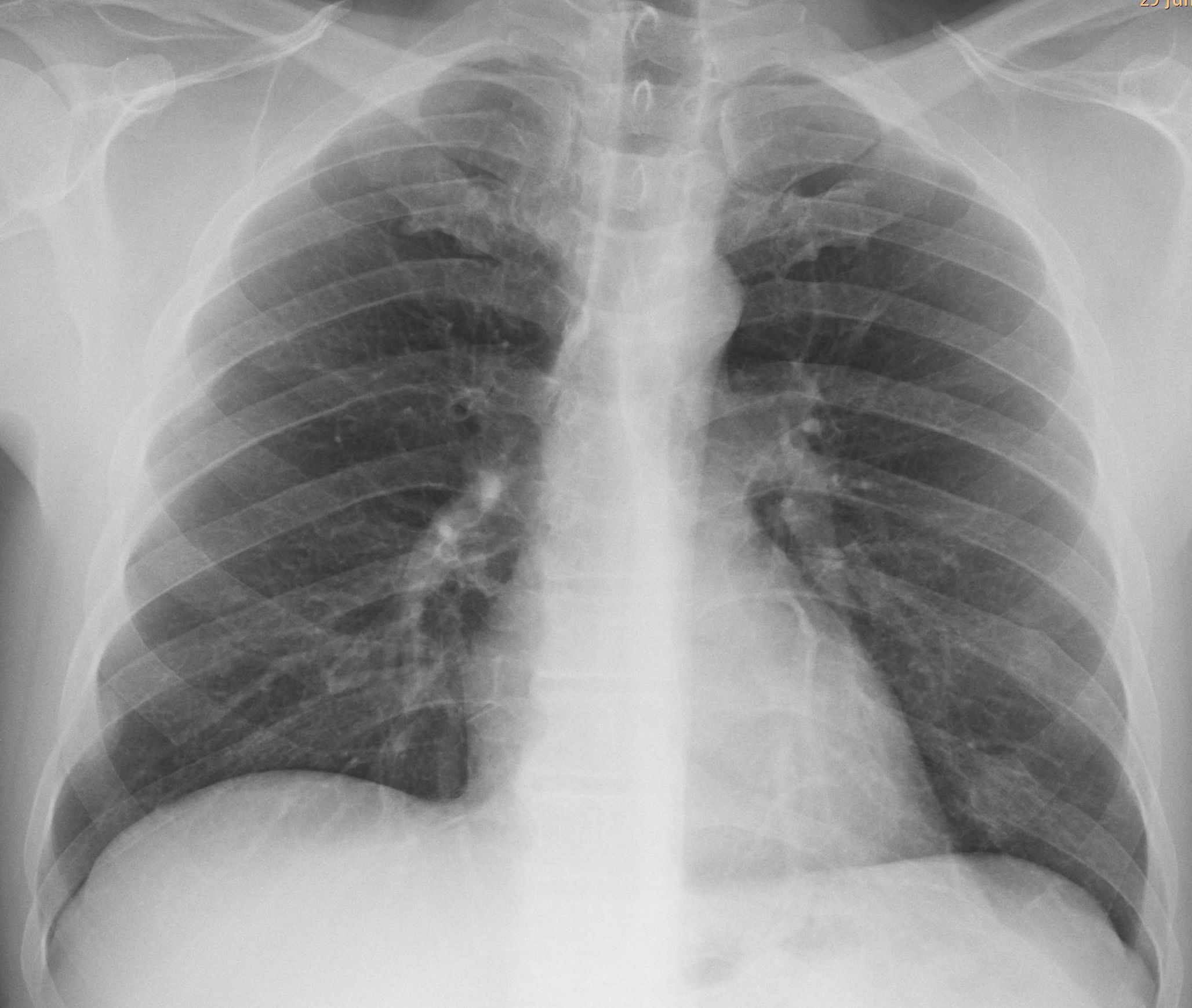
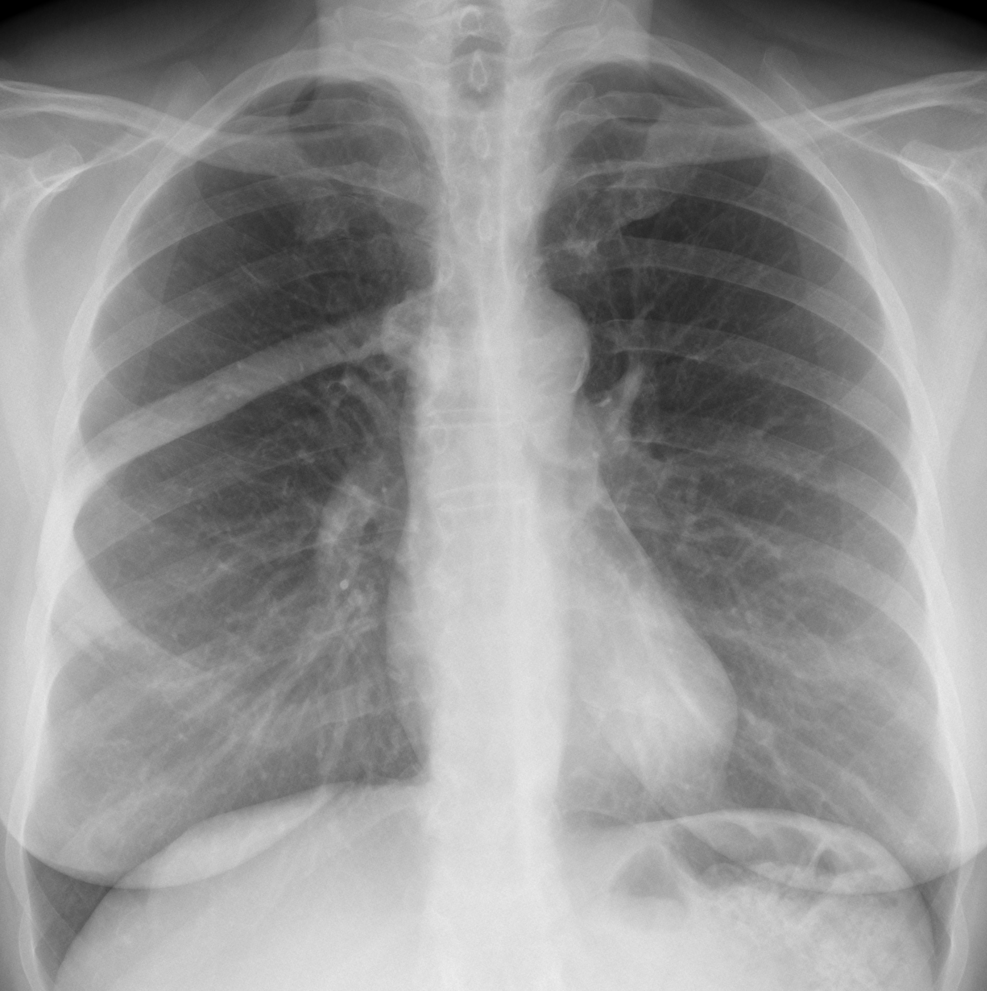
I am doing spring cleaning of my cases and this week I am showing two of them. Radiographs belong to two unrelated middle-age patients with chest pain.
What do you see?
Check the images below, leave your thoughts in the comments section, and come back on Friday for the answer.
Case 1 findings: PA radiograph shows an extrapulmonary lesion at the periphery of the right hemithorax (A,B arrows). Those of you that diagnosed a rib fracture are very perceptive, because I did not see it.
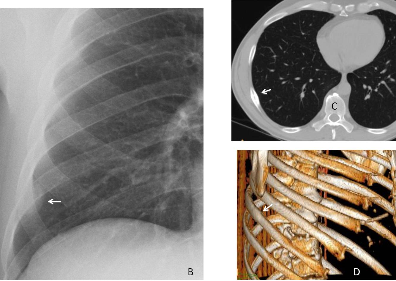
The patient was recalled and a CT showed disappearance of the extrapleural haematoma and the callus from the healed fracture (C-D, arrows).
Final diagnosis: acute rib fracture with extrapleural hematoma
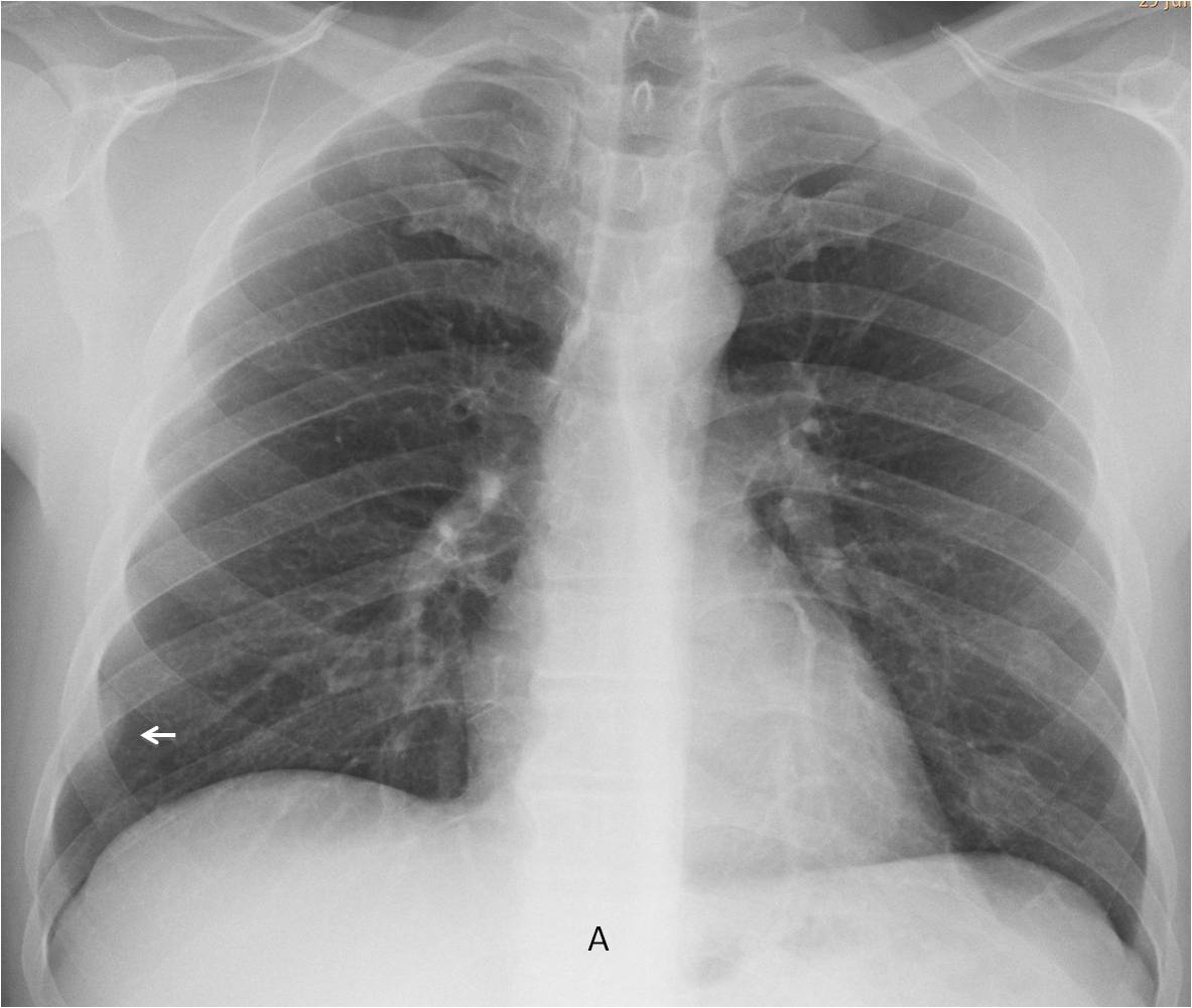
Case 2 findings: Chest radiograph shows a sclerotic right 6th rib (E, arrow). The appearance of the rib suggests a benign lesion, supported by a previous report five years earlier (no radiograph, sorry) in which the sclerotic rib was described.
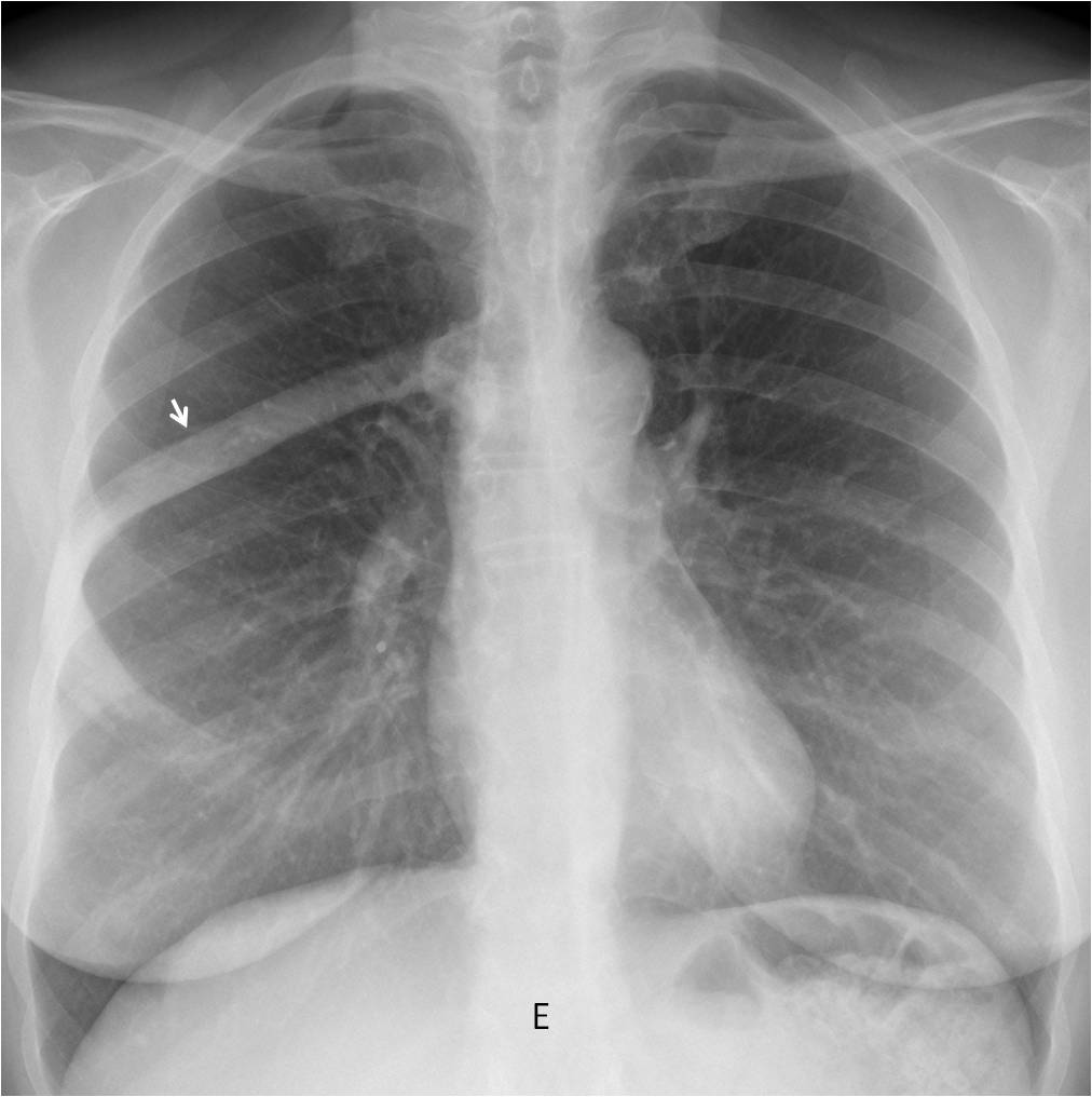
In my opinion, the facts that the rib is enlargedand and the sclerosis reaches the distal ends, and the thickened cortical, point to Paget disease.
Final diagnosis: probably Paget disease of the 6th rib (unproven)
Congratulations to the majority of you who suggested the right diagnosis. To me you are all winners.
Teaching point: fracture is the most common rib lesion. It should be our first diagnosis even if the findings are not clear, as you proved in this case.