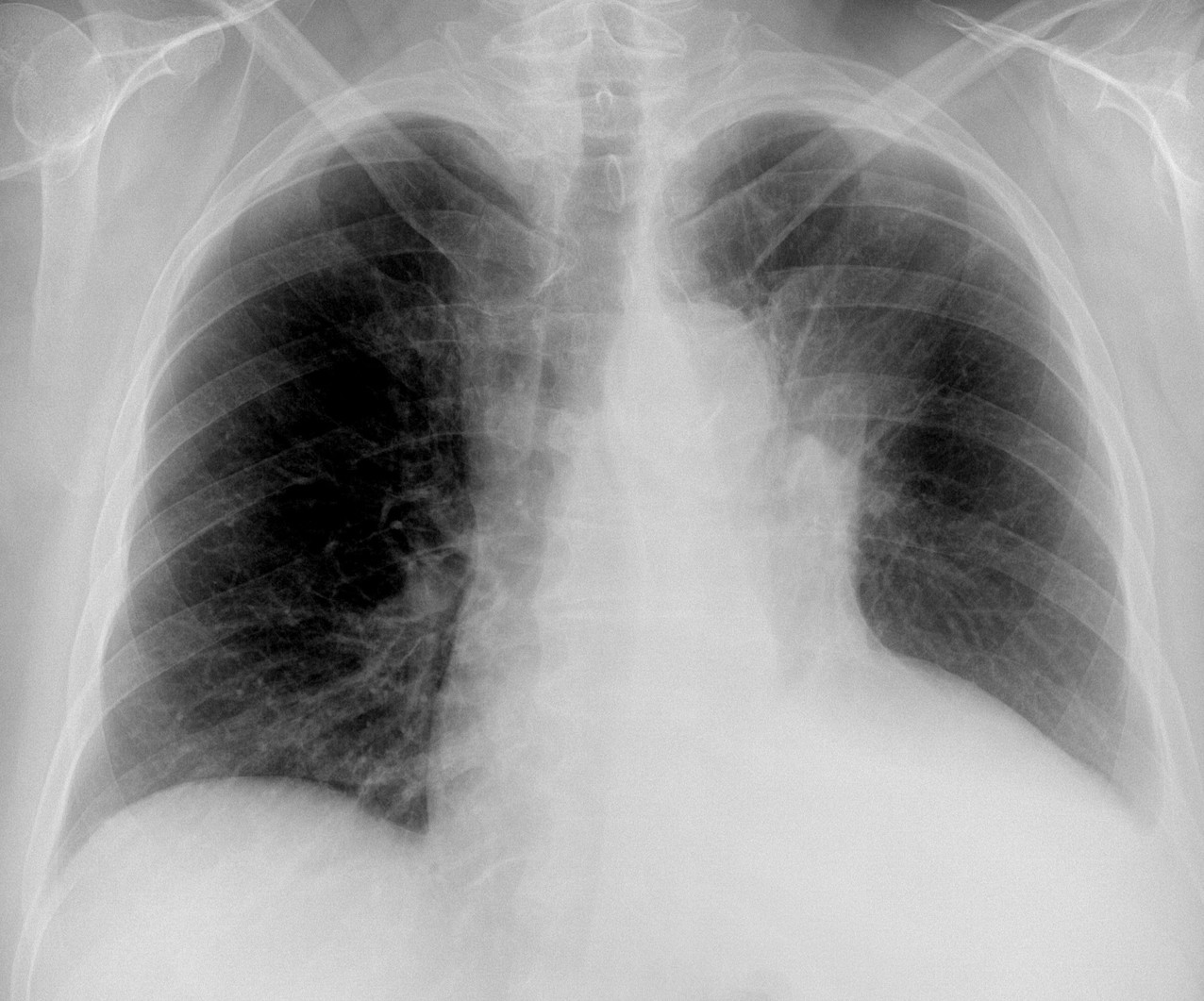
Today I want to test your prowess in plain film interpretation. The PA radiograph belongs to a 79-year-old woman with a cough and haemoptysis. What do you see?
Check the image below, leave your thoughts in the comments section, and come back on Friday for the answer.
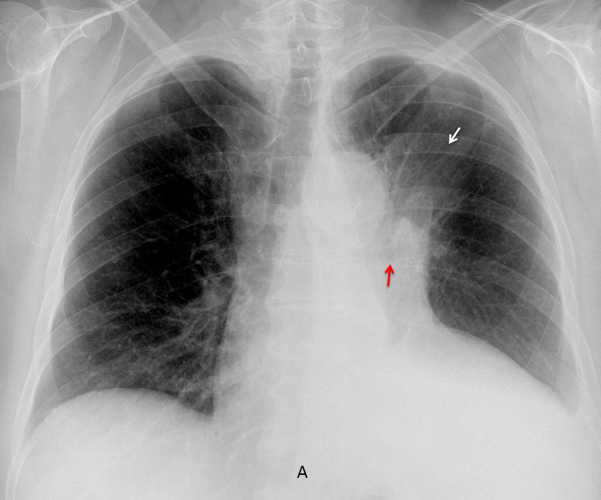
Findings: PA chest radiograph shows an ill-defined pulmonary opacity in the left upper lung (A, arrow). The left hilum is elevated and the left main bronchus is arched upward (A, red arrow). This combination of findings is highly suggestive of LUL collapse. There is also blunting of the left costophrenic angle, indicative of pleural fluid and apparent cardiomegaly.
Enhanced CT confirms the LUL collapse (B, arrow), due to a mass at the origin of the LUL bronchus (C, arrow). An interesting finding is that the heart is of normal size (B-D, red arrows).
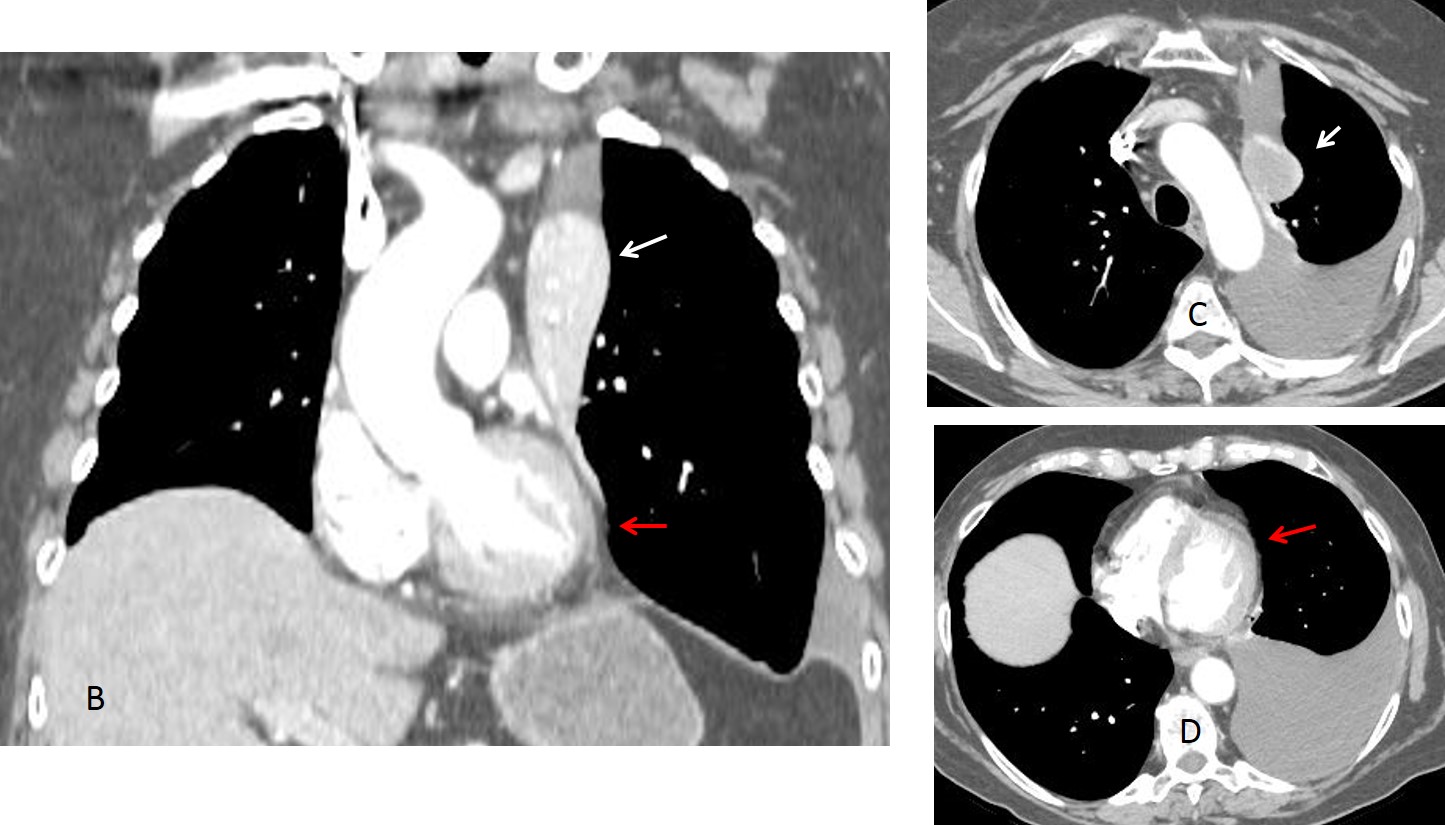
The apparent cardiomegaly is due to the apposition of pleural fluid over the cardiac shadow in the upright chest radiograph. This happens occasionally and you can see two examples in Diploma case 85. I don’t know if this has been reported in the literature.
Final diagnosis: carcinoma with collapse of LUL and pleural effusion simulating cardiomegaly.
Congratulations to TR, who was the first to suggest the diagnosis, although the rest of you did very well too.
Teaching point: it is important to recognise the signs of LUL collapse in the PA view, because carcinoma is the most common etiology. An added bonus in this case is remembering that left pleural effusion may simulate cardiomegaly in the upright PA view.