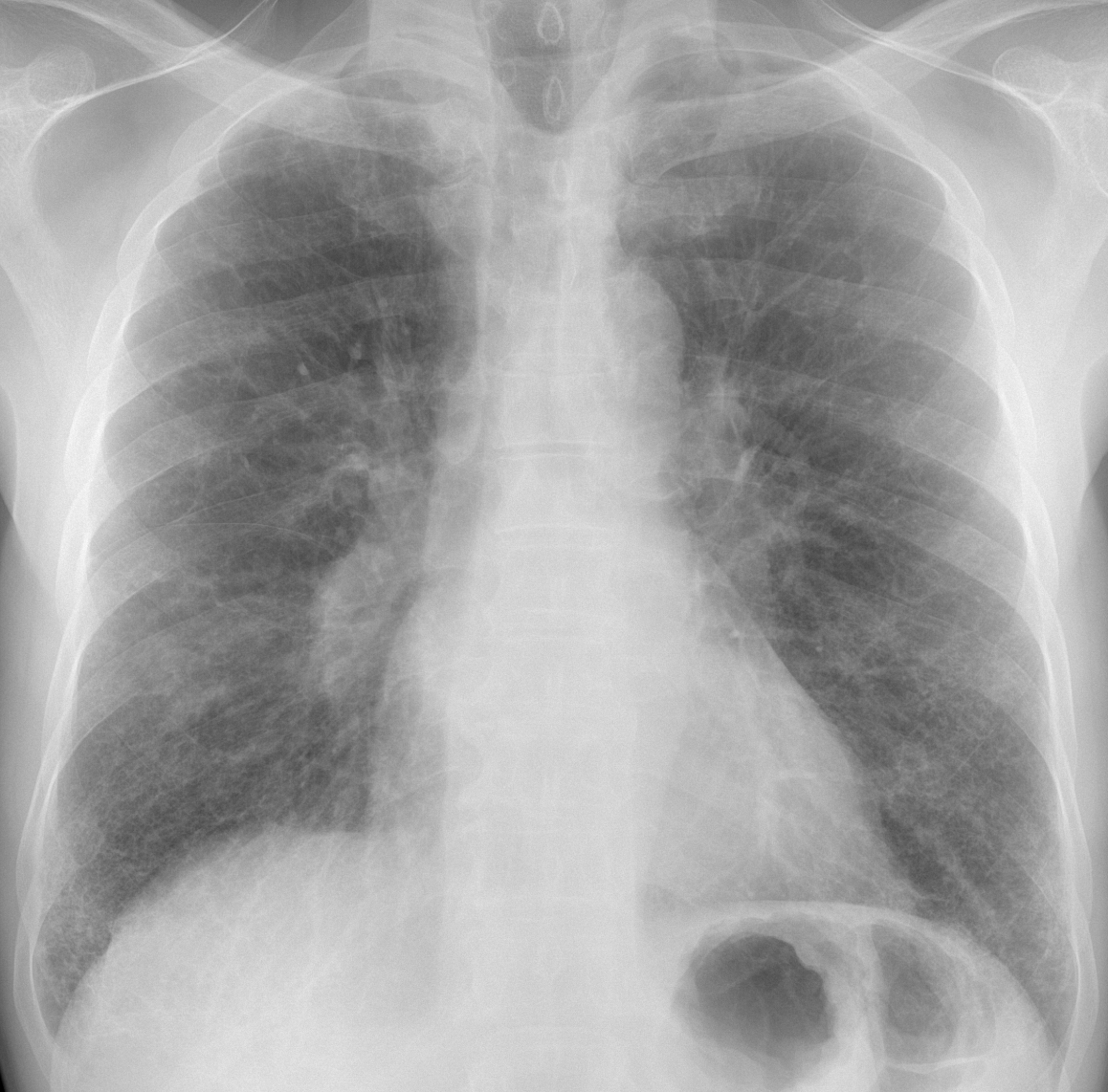
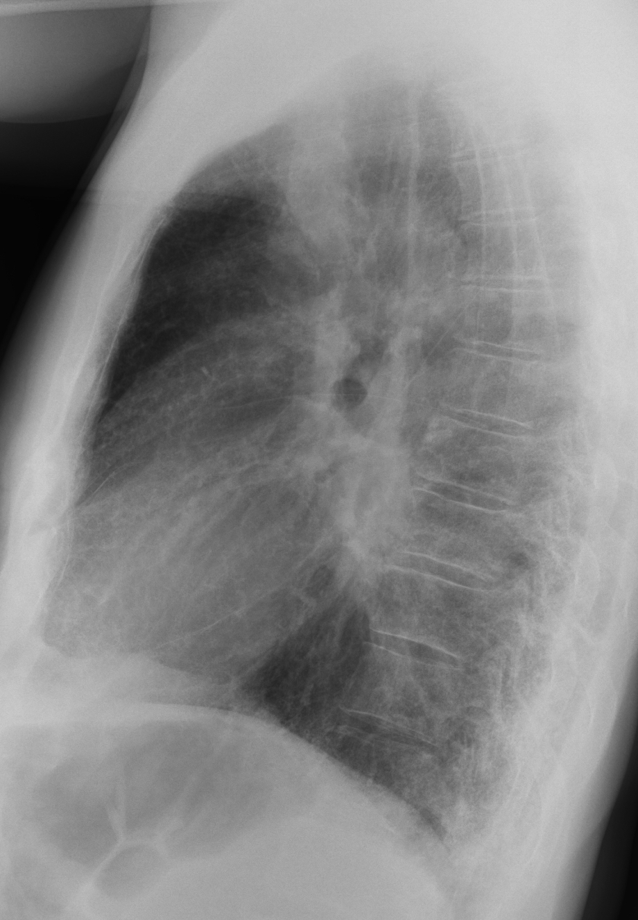
Today I am showing a case seen one month ago. Radiographs belong to a 64-year-old man with intermittent fever for the previous two weeks. What do you see?
Check the images below, leave your thoughts in the comments section and come back on Friday for the answer.
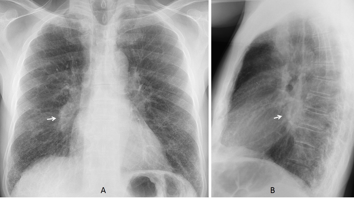
Findings: in the PA view there is a fine interstitial pattern in both lung bases. I believe that the rest of the lung is normal. What caught our attention was the increased opacity of the lower right hilum, visible in the PA and lateral projections (A-B, arrows).
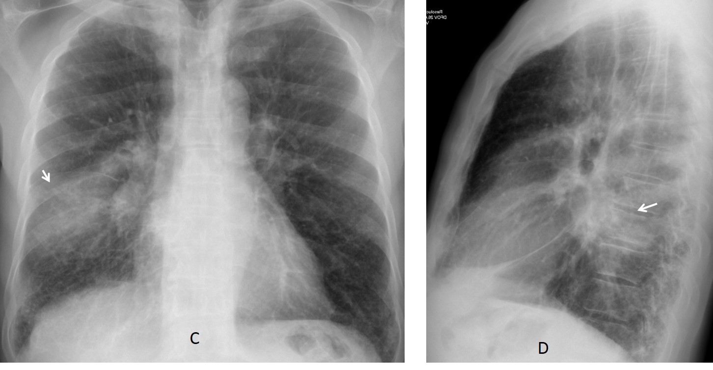
We suspected unilateral enlarged lymph nodes and recommended CT. The attending physician did not follow the recommendation until the patient returned three weeks later with high fever and an infiltrate in the right lung (C-D, arrows).
Enhanced CT confirms the basal fibrosis (E) as well as a large PET positive mass in mediastinum and right hilum (F-G, arrows), better seen in the coronal and sagittal reconstructions (H-I, arrows). Biopsy confirmed a squamous-cell carcinoma.
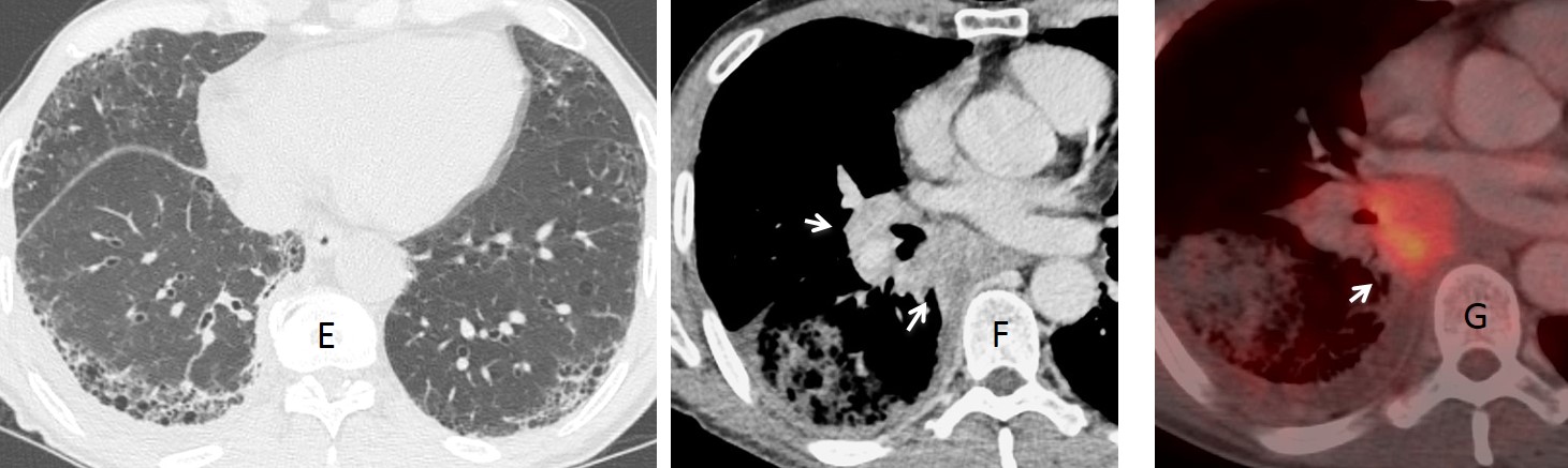
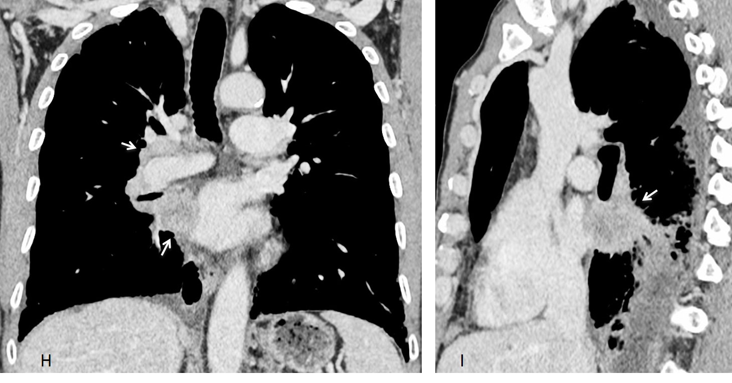
Final diagnosis: Squamous-cell carcinoma of right hilum and mediastinum, barely visible in the chest radiograph. Basal pulmonary fibrosis.
This was a difficult case and I believe most of you gave your best. I must single out Olena, because she described the alterations in the right hilum and was very perseverant.
Teaching point: avoid satisfaction of search and remember to look at the lateral view.