Today I want to try a new way of presenting cases: conventional PA and lateral radiographs will be shown on Monday, followed by additional images on Wednesday. Hope this approach will have more educational value. If it works well, I will do it more often.
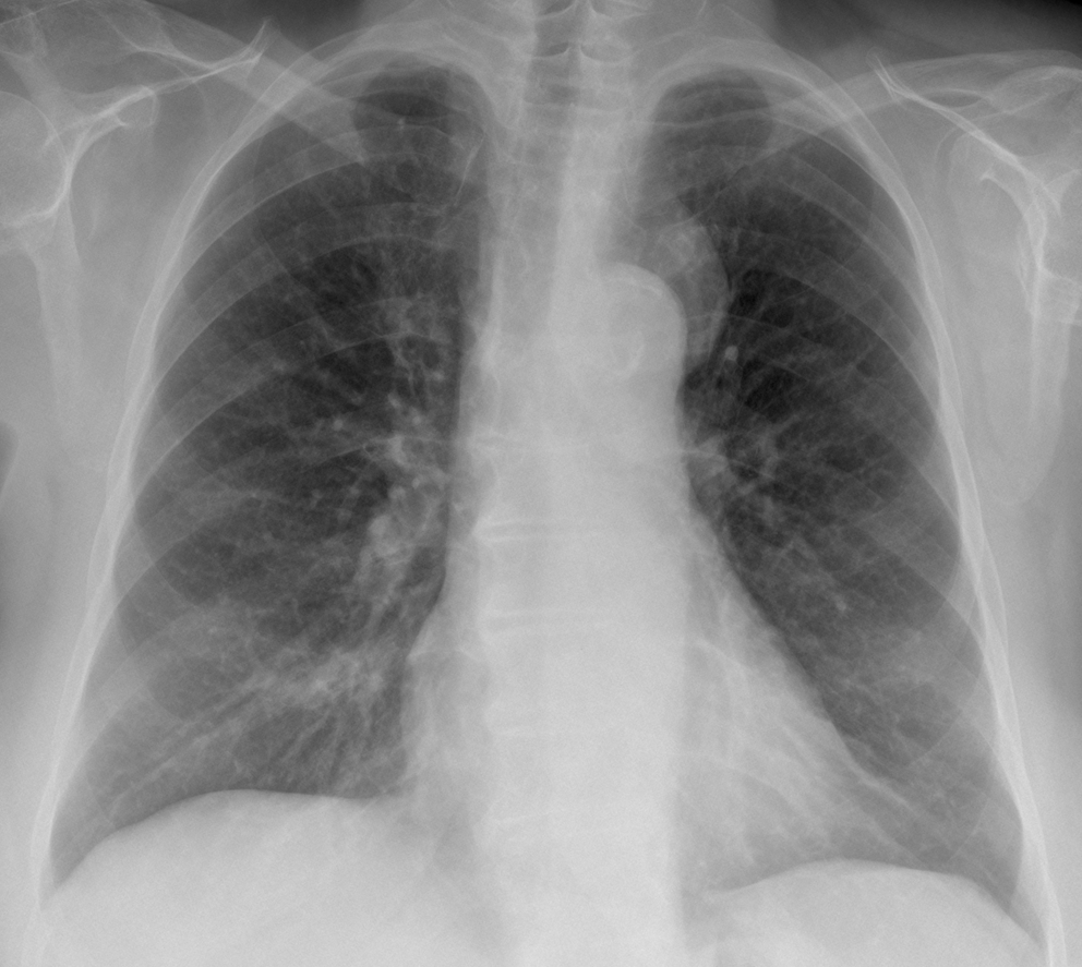
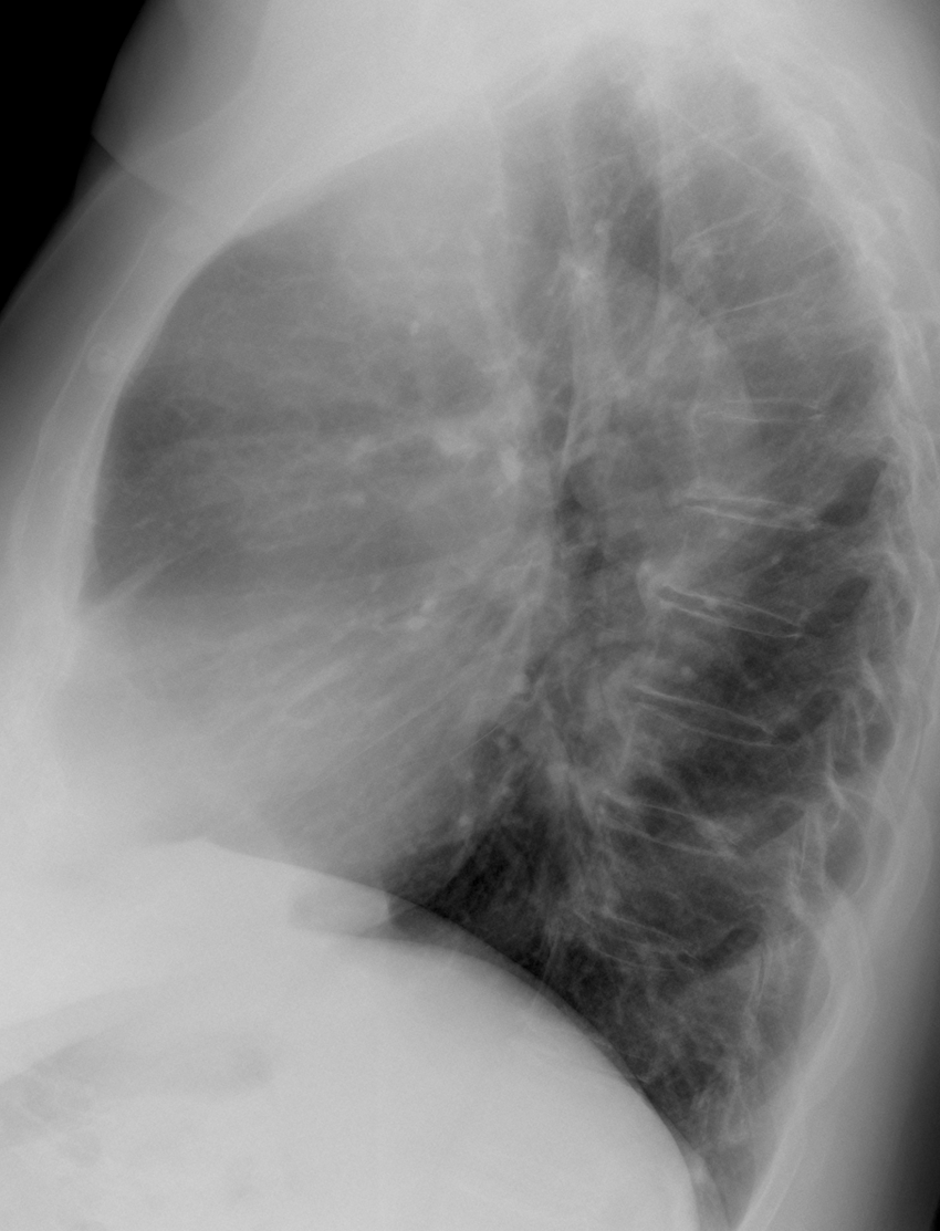
Today’s radiographs belong to a 74-year-old woman with mild respiratory infection.
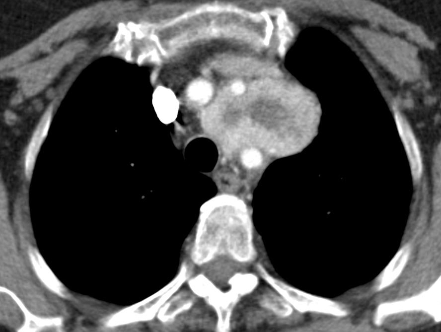
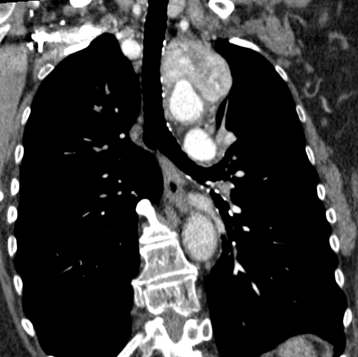
As promised, here are two additional images of the enhanced CT. Are they of any help?
Findings: chest radiographs show a superior/anterior mediastinal mass projected over the aortic knob (A-B, arrows).
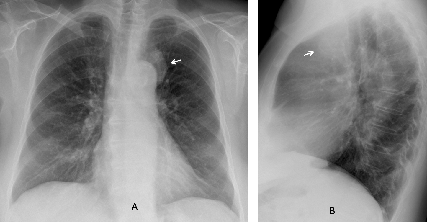
The classic differential diagnosis of a mass in this location lies among thymoma, embriogenic tumour and lymphoma (less likely because of the paucity of symptoms). Intrathoracic goiter would be unusual in front of the trachea.
Enhanced coronal and axial CT show an intensely enhancing mass with necrotic center located between the supra-aortic vessels (C-D, arrows).
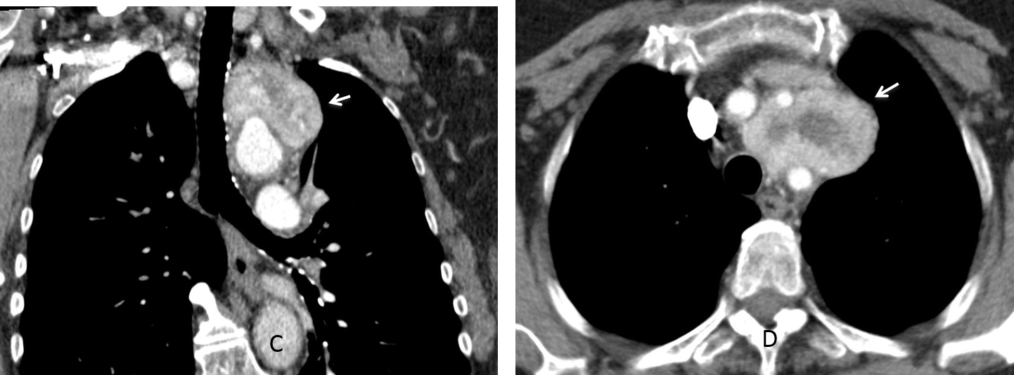
The differential diagnosis of avid enhancing mediastinal masses includes thyroid, paragangliomas and Castleman disease. In this particular case, the location between the vessels favors the diagnosis of paraganglioma.
SPECT-CT with (111)In octeotride shows marked uptake of the mass (E-F, arrows).
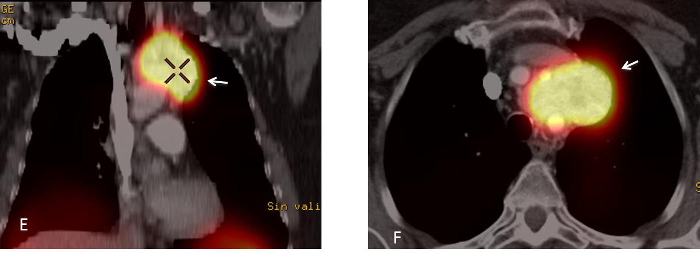
Final diagnosis: paraganglioma of upper mediastinum.
Contratulations to TR, winner of the first phase, and MK who made the correct diagnosis.
Teaching point: Remember that an avidly enhancing mass in the mediastinum, suggests endothoracic goiter as the first possibility, followed by paraganglioma and Castleman disease.
My heartfelt thanks to Dr. Eva Castañer, who provided the CT and SPECT images.