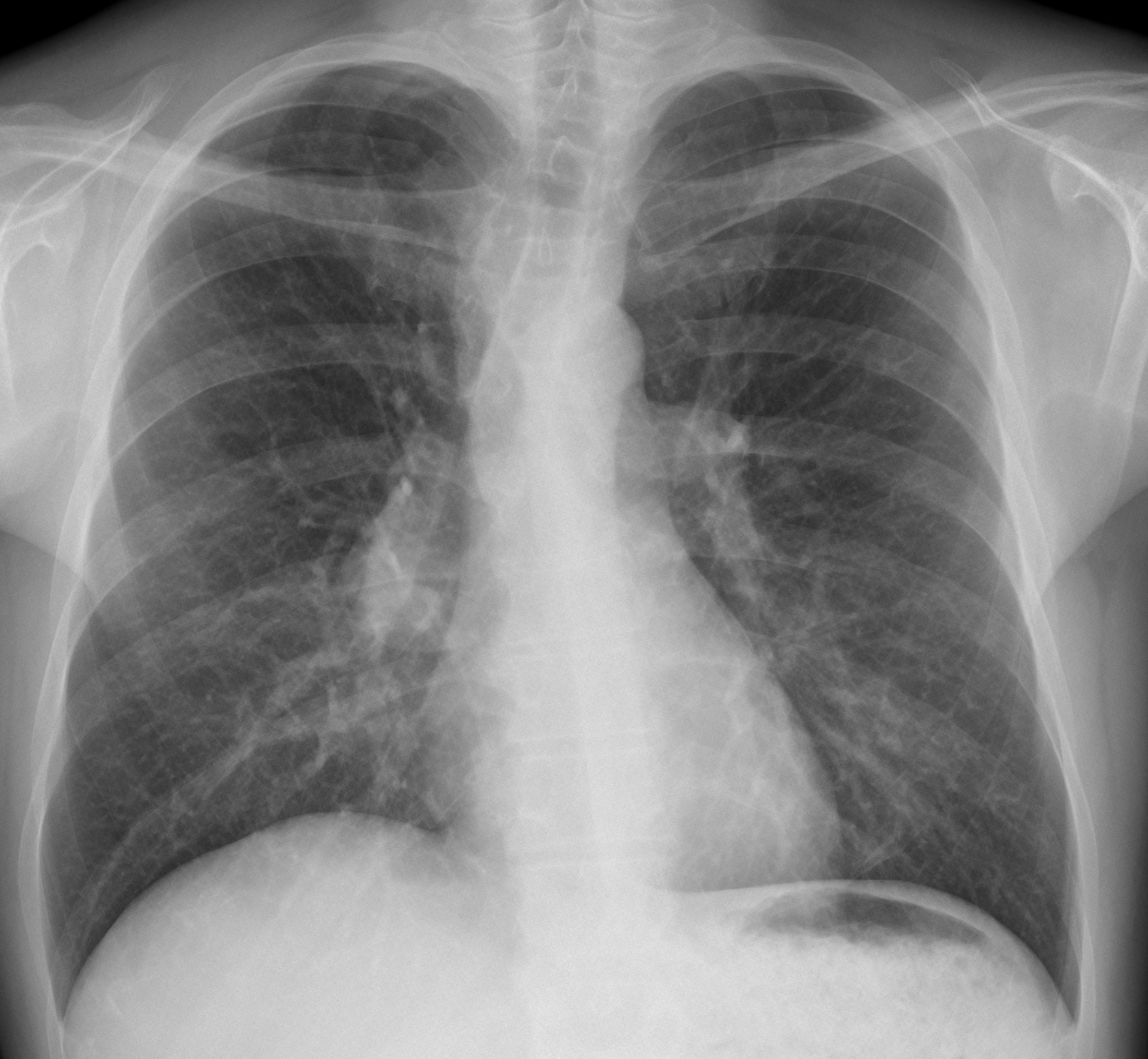
Today I am showing a PA radiograph of a 48-year-old smoker with a persistent cough. What do you see?
As in last week’s case, I will show additional images on Wednesday morning.
Check the images below, leave your thoughts in the comments section and come back on Friday for the answer.
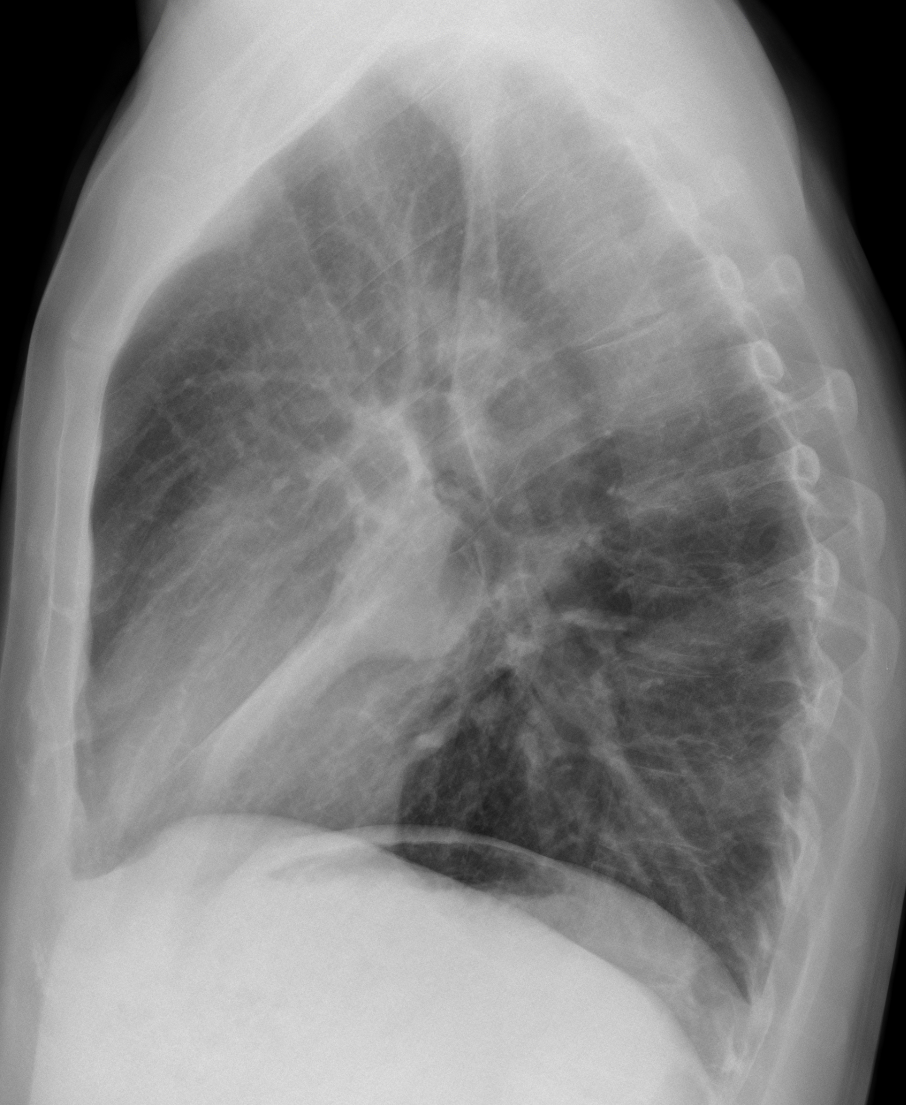
As promised, here are the two additional images. Do they help you to reach a conclusion?
Findings: PA radiograph shows a well-defined nodular shadow superimposed to the right hilum (A, arrows). Lateral view shows a rounded mass in the right hilum (B, arrow), causing marked collapse of the RML (B, red arrow).

Sagittal CT confirms the mass (C, arrow) and the RML collapse (C, red arrow).
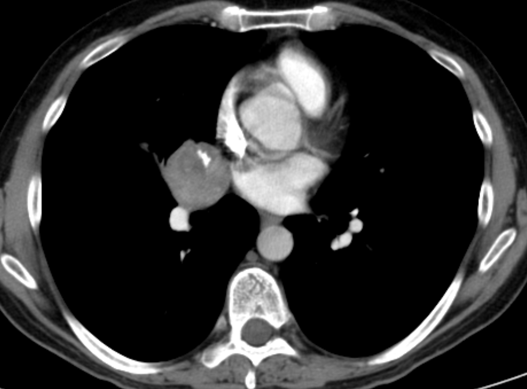
There is coarse calcification within the mass (C-D, yellow arrows). PET-CT shows marked uptake of the mass (E, arrow).
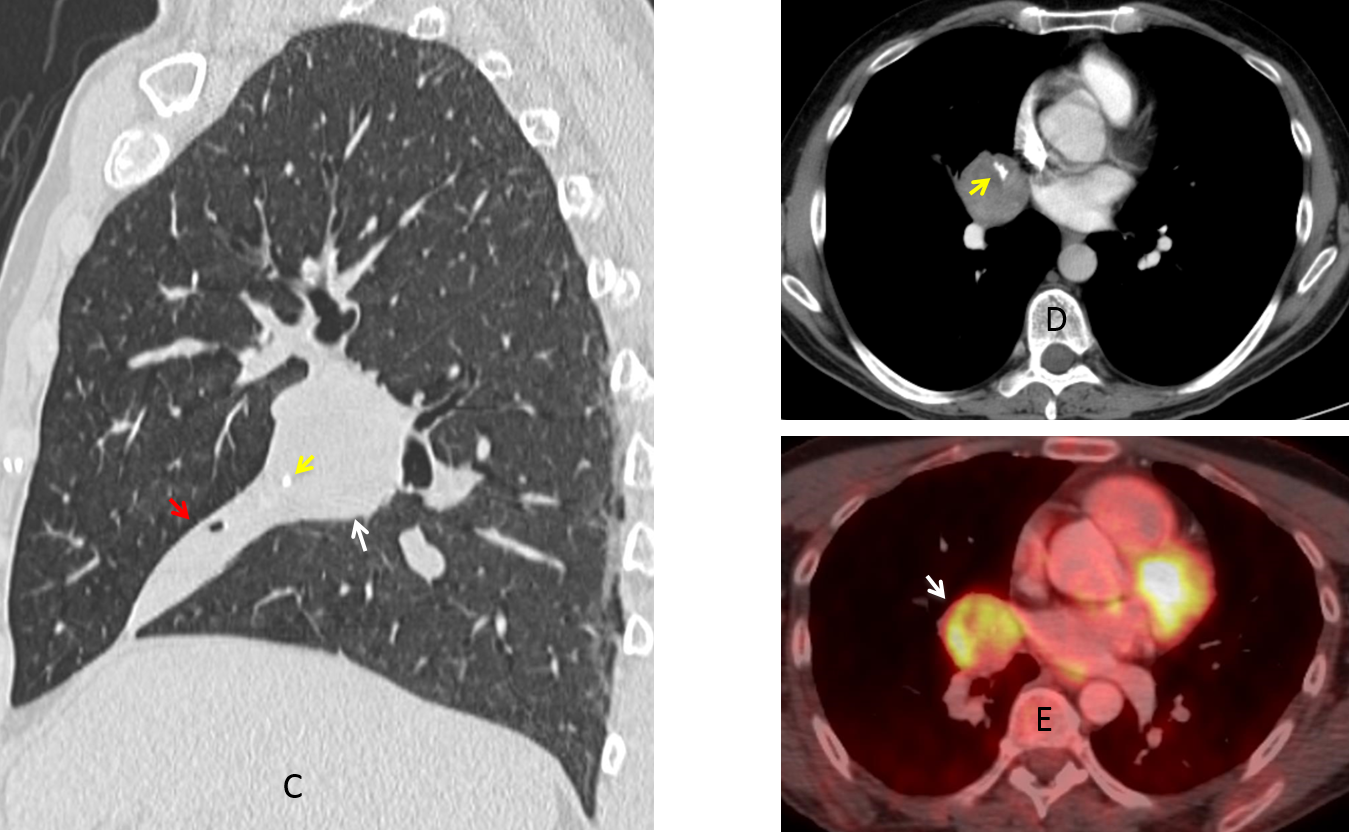
Bronchoscopy confirmed a mass occluding the RML bronchus. Biopsy returned the diagnosis of carcinoid.
Final diagnosis: carcinoid tumor causing marked collapse of RML.
Congratulations to MK, who was the first to mention the correct diagnosis.
Teaching point: this case confirms the importance of the lateral view to complement the information obtained in the PA radiograph. And remember that a nodule with coarse calcification in CT practically excludes bronchogenic carcinoma.