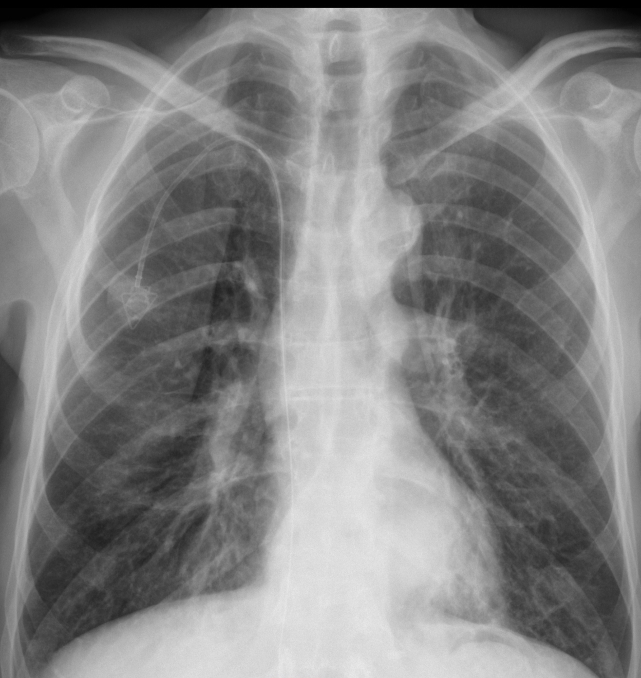
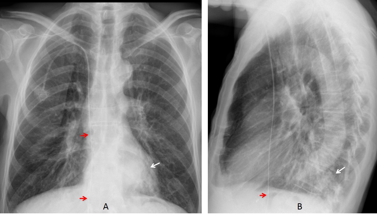
Case 1 findings: PA chest radiograph shows a retrocardiac pulmonary infiltrate (A, arrow), confirmed in the lateral view (B, arrow). Although the patient did not have significant symptoms, it was considered to be inflammatory and cleared with antibiotics. An additional finding is the path of the guiding wire of the port-a-cath, which is clearly going beyond the right atrium into the abdomen (A-B, red arrows).
CT scout view after removal of the port-a-cath shows the wire remaining in the SVC, with the distal end in the left iliac vein (C, arrows). Coronal CT taken four years later shows no change (D, arrows).
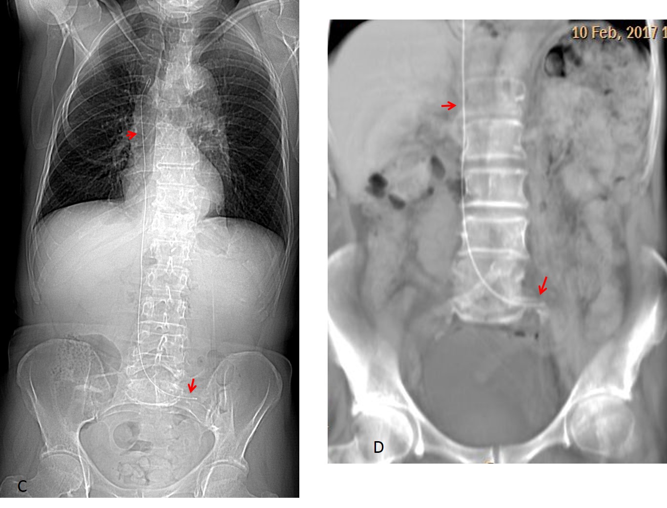
Final diagnosis: LL pneumonia. Wire lodged in SVC and IVC.
Case 2 findings: PA chest radiograph shows fibrotic changes in the right apex and a faint nodule in the mid left lung fiend (A, arrow), not visible one year earlier (B, circle), raising the possibility of a metastatic nodule.

Axial CT and 3-D reconstruction show that the nodule represents a healed fracture of the anterior fifth rib (C-D, arrows) accompanied by another fracture of the sixth rib (D, yellow arrow).

Final diagnosis: unsuspected rib fracture simulating a metastatic nodule.
Congratulations to LAR, who saw the abnormal path of the port-a-cath and to Coffee, who was the first to detect the nodule if the left lung.
Teaching point: I selected these two cases to cover for Dr. Pepe, thinking that the diagnosis would be easy. Remember the KISS method!