Today’s images belong to a 38-year-old woman treated with antibiotics for a sore throat with fever. Six days later she goes to the ER with left chest pain and moderate dyspnea.
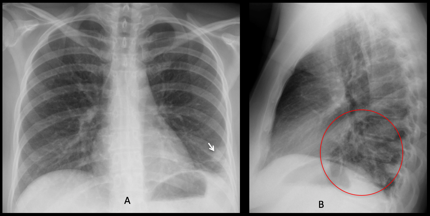
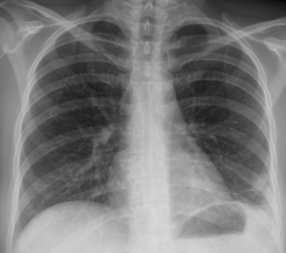
Findings: PA chest radiograph shows a pulmonary opacity at the left lung base (A, arrow), with obliteration of the costophrenic angle. The opacity is also visible in the lateral view (B, circle). Its appearance is non-specific, although the location favors a pulmonary infarct.
In this case, the main diagnostic help is the clinical information: chest pain and pulmonary infiltrates associated to a recent oropharyngeal infection suggest internal jugular thrombosis with associated sepsis and pulmonary emboli (Lemierre syndrome).
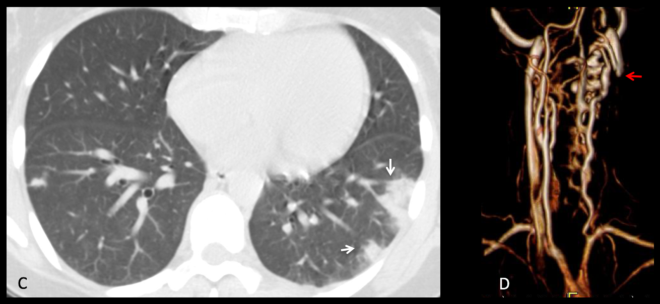
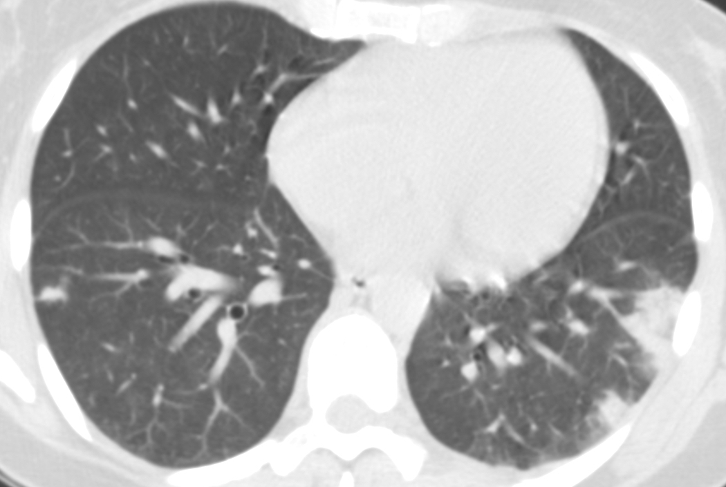
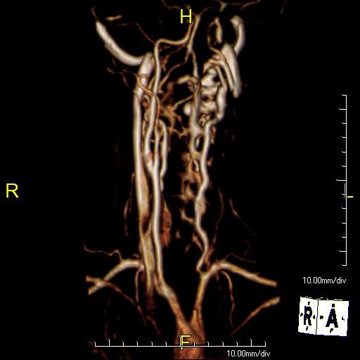
Axial CT shows left basal infiltrates suggestive of pulmonary infarcts (C, arrows). Doppler study and MR angio confirm thrombosis of the left jugular vein (D, red arrow). Blood culture was positive for Fusobacterium necrophorum.
Final diagnosis: Lemierre syndrome.
Congratulations to MK, who made an early diagnosis.
Teaching point: although Lemierre syndrome is supposed to be a rare clinical entity, the radiologist can suggest the diagnosis if she/he knows the clinical presentation, prompting early treatment.


I see a faint rounded soft tissue density in the left lower lung zone which seems to be arising from the chest wall. I do not see any associated fractures or rib destruction and the border does not seem completely defined to me. There may also be some linear atelectasis in this region.
Considering her chest pain and dyspnea I would consider a pleural fibroma.
There is focal opacity with bulging contour and few reticular opacity at periphery of left lower lung zone with blunting left costophrenic angle, seem to be eliptical shape or more elongated in lateral view, possibly pleural lesion such as pleuritis with loculated pleural effusion or empyema.
Hello! There is an obliteration of the left costophrenic angle and a well defined internal margin of a lesion that can be pulmonary or extrapleural (I think I need a CT to define the localization). The hystory remembers me a Lemierre syndrome with pulmonary septic embolism (in this case the lesion will be intrapulmonary ). Any signs of yugular thrombophlebitis vein?
More images will be shown next Wednesday
Alveolar periferic infiltrates (bigger in size and number in LLL-septic embolims).
At vascular reconstruction the left yugular vein is not visible so a thrombosis have to be suspected.
Greetings,
peripheral incompletely round opacity over the left lower zone that cannot be seen on the lateral view, the lesion is probably related to the chest wall/breast, and the haziness over the left lateral costophrenic recess, I think, can be explained by perilesional soft tissue reaction/edema.
Greetings,
I am not so sure about what I see in the angiogram, the left common carotid looks blocked shortly after its origin, which I think explains the nonvisualization of the left jugular vein.
Peripheral pleural based opacities in the left lung suggesting embolic event.
I could not figure outvthe connection!
The flow in the left carotid was OK. The left jugular was thrombosed.
I didn’t select the best image. Sorry.
Greetings,
in this case I suggest leimierre syndrome.
Greetings,
may I ask why the neck vessels angiogram was done?, was there any symptoms in the neck?.
thank you
Remember that the patient originally had a sore throat. After the chest CT a Lemierre sindrome was suspected and a Doppler study confirmed thrombosis of the jugular vein. Personally, I think the MR study was unnecessary, but it was done, anyway.