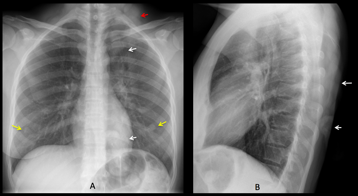
Findings: PA radiograph shows an ill-defined opacity in the left lung, running from top to bottom (A, white arrows). The opacity extends towards the neck (A, red arrow), which suggests that it is external to the lung. There are two nodules in the lower lungs that correspond to the nipples (A, yellow arrows). Lateral view shows an elongated opacity in the back of the chest (B, arrows).

A photo of the patient (C) confirms that a long braid is the cause of the opacity. PA radiograph after lifting the braid shows a normal chest (D).
Final diagnosis: hair braid simulating a pulmonary infiltrate.
Congratulations to MK, who was the first to suggest the diagnosis.
Teaching point: remember that long hair may project over the lung, simulating pulmonary nodules or infiltrates. The clue to the diagnosis lies in recognizing that the abnormality extends to the neck.
Good morning! There are bilateral nodular opacity at lower location (I think they are the nipples). There is an increased density proyected over the supraclavicular left region, upper left lobe, over the hilum and in retrocardiac location. I would like to see the lateral view and go to see the patient and its hair…… Long pigtail?
Oh yes! No left breast shadow…
No breast shadow and a floating nipple?
Curioser and curioser…
Hello. I don´t see the left mammary shadow (previous op?). There is a small bulge in the contour of the right upper mediastinum (rt aortic arch?). I can also see the density on the left, as mentioned by MK, that begins in the level of the supraclavicular soft tissues and extends down at least to the level of the hilum.
Assymetrical breast shadow, preserving both nipple opacities (anatomical asymmetry? breast conservating surgery?)
Increased density in left apex and supraclavicular area, extending to retrocardiac/diaphragmatic location, as commented by MK.
Ill defined nodular opacity (I can only delineate its lower margin, located lateral to the right hilum (pulmonary nodule?)
left mastectomy – was the endometrial cancer linked to tamoxifen?
hair braids simulating opacity in left supraclavcular region going to left hilum.
Mass behind the heart on the left extending below the diaphragm – unsure. Compare with previous old CXR. ?? sequested lung vs a malignant mass. I would CT.
The left breast is present, although flattened. The nipple is visible at the left base.
You are right about the hair braid. It goes all the way down and simulates the retrocardiac mass. See the answer later today