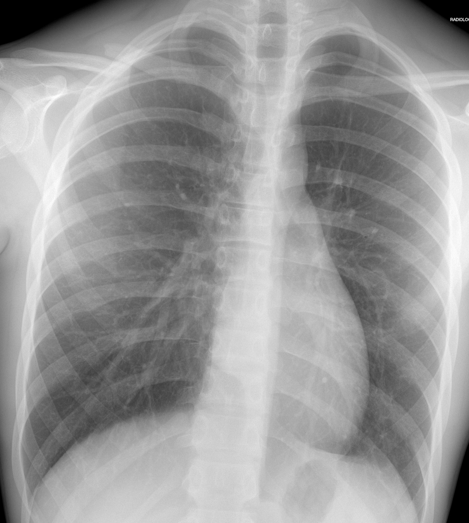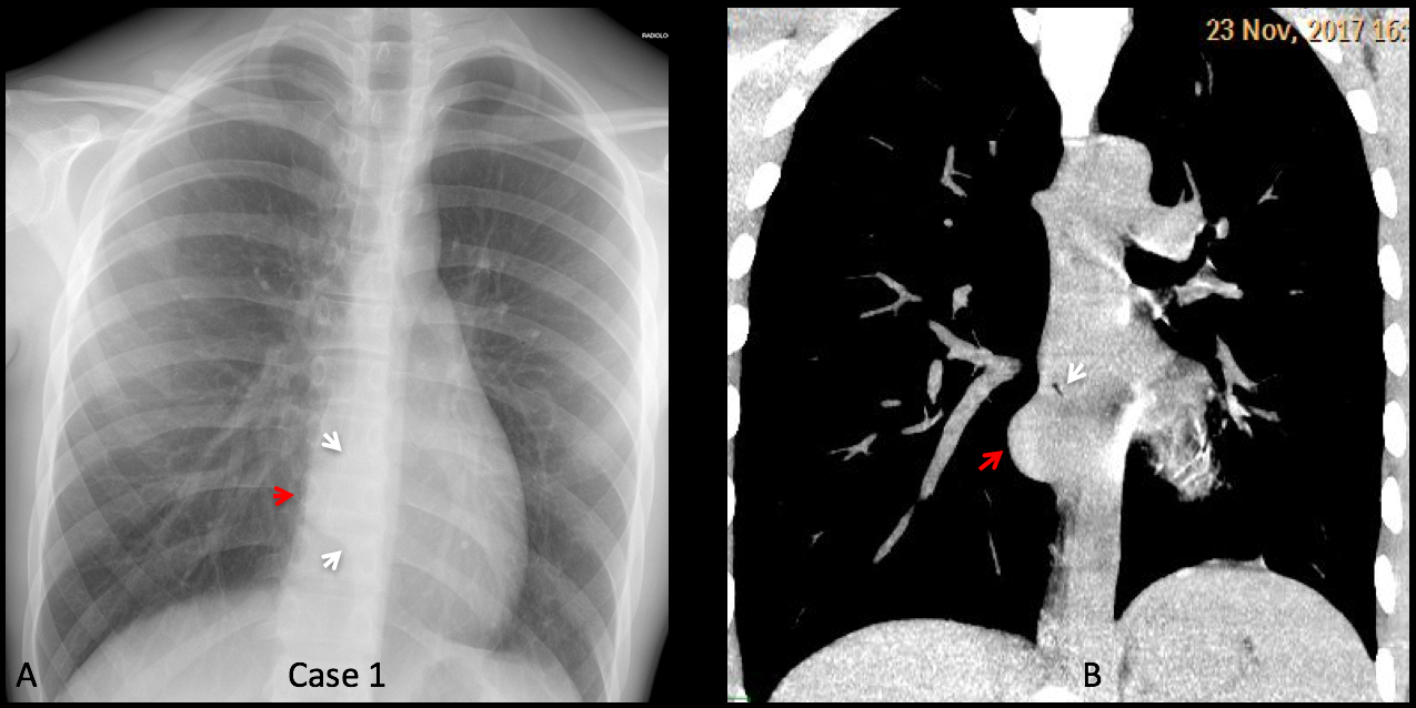
Findings case 1: there is an interruption of the para-esophageal line (A, arrows) by a rounded middle mediastinal opacity (A, red arrow). In a young asymptomatic patient, the most probable diagnosis is an esophageal duplication cyst.
Unenhanced coronal CT confirms the mass (B, red arrow) adjacent to esophageal air (B, arrow). The density of the mas is similar to the mediastinal soft tissues, which is not uncommon in duplication cysts. To confirm the diagnosis an MRI was ordered. Unfortunately, this week in Spain has two holidays (very few of us are working) and the MRI will not be done until next week.
Diagnosis: probable mediastinal duplication cyst, pending MRI.
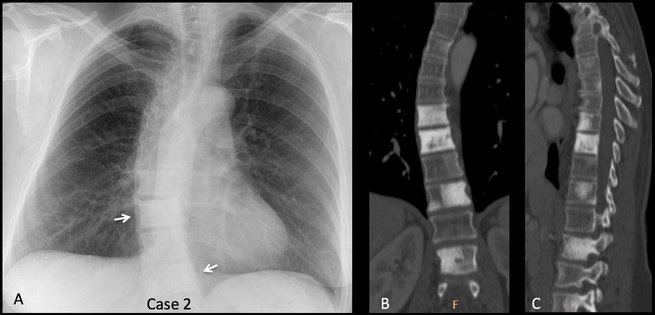
Findings case 2: there is moderate scoliosis and sclerotic vertebras are visible through the mediastinal shadow (A, arrows).
Coronal and sagittal CT (B, C) confirms the findings. Review of the clinical history discovered that the patient had a previous carcinoma of the breast.
Diagnosis: sclerotic vertebral metastasis from breast carcinoma.
Congratulations to Coffee, who was the first to diagnose both cases.
Teaching point: these are two nice cases, seen the same week, in which the abnormalities were inside the mediastinal shadow and could be easily missed (as a matter of fact, two of you did).