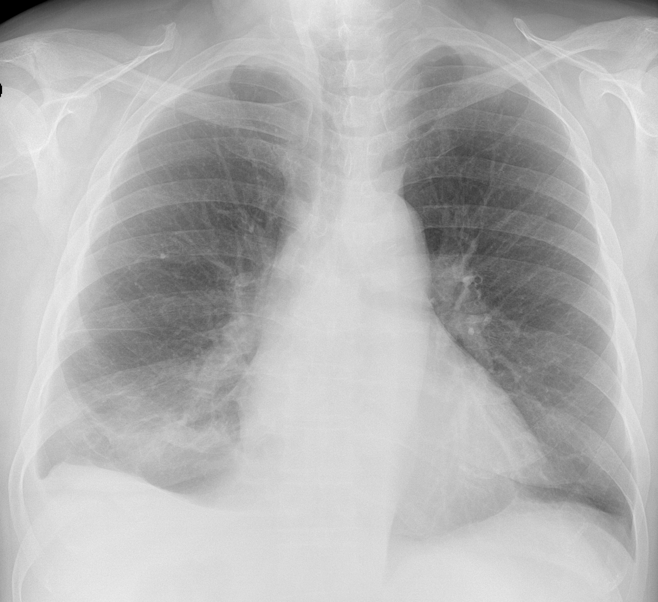
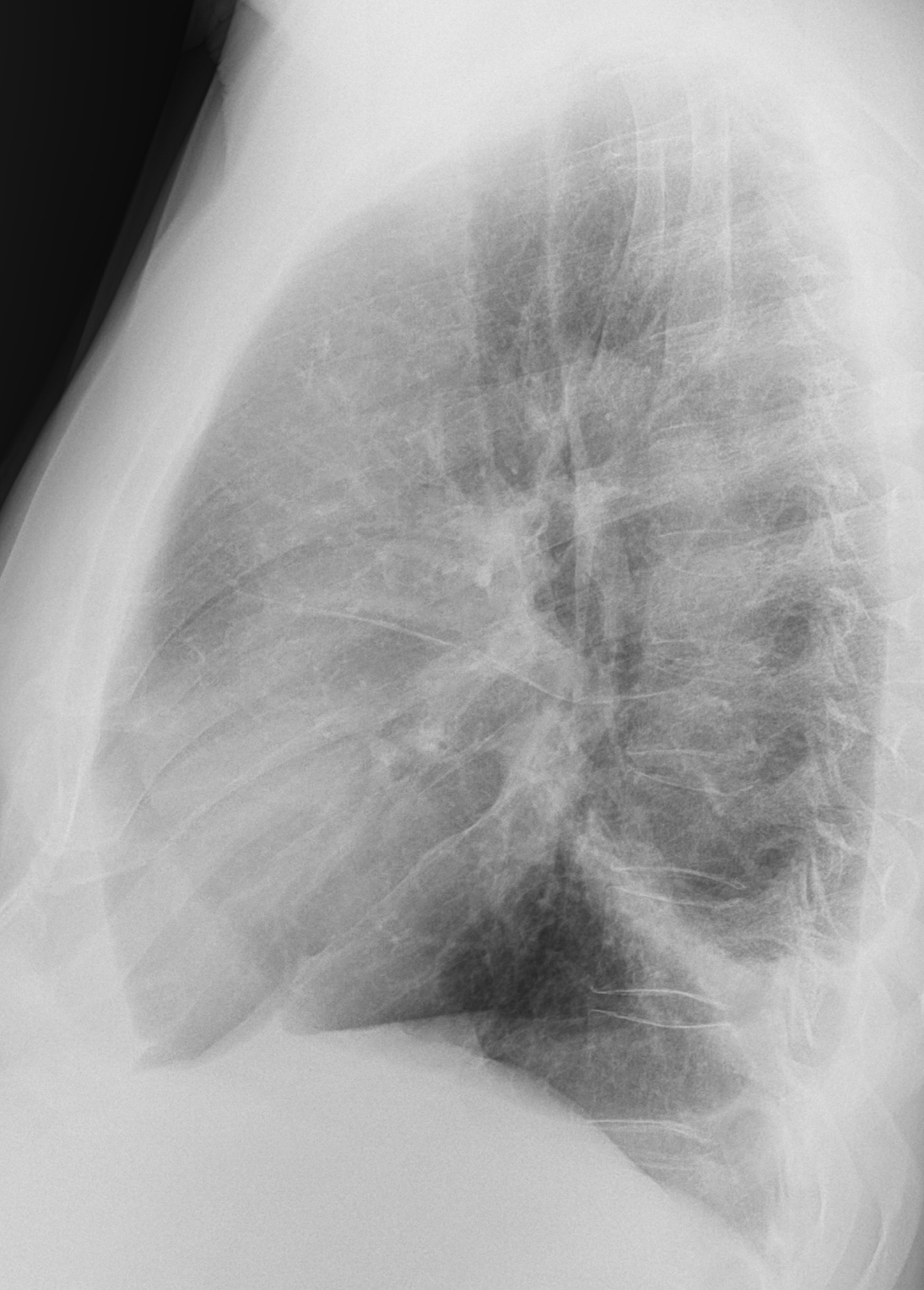
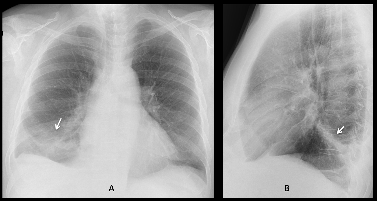
Findings: the PA radiograph shows an ill-defined opacity in the RLL (A, arrow), accompanied by a small pleural effusion. The lateral view shows involvement of the posterior segment of the RLL (B, arrow).
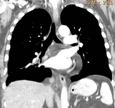
Sagittal CT confirms the segmental involvement (C, arrow). Coronal and sagittal CT with mediastinal window shows a low-density lesion at the origin of the bronchus (D-E, red arrows).
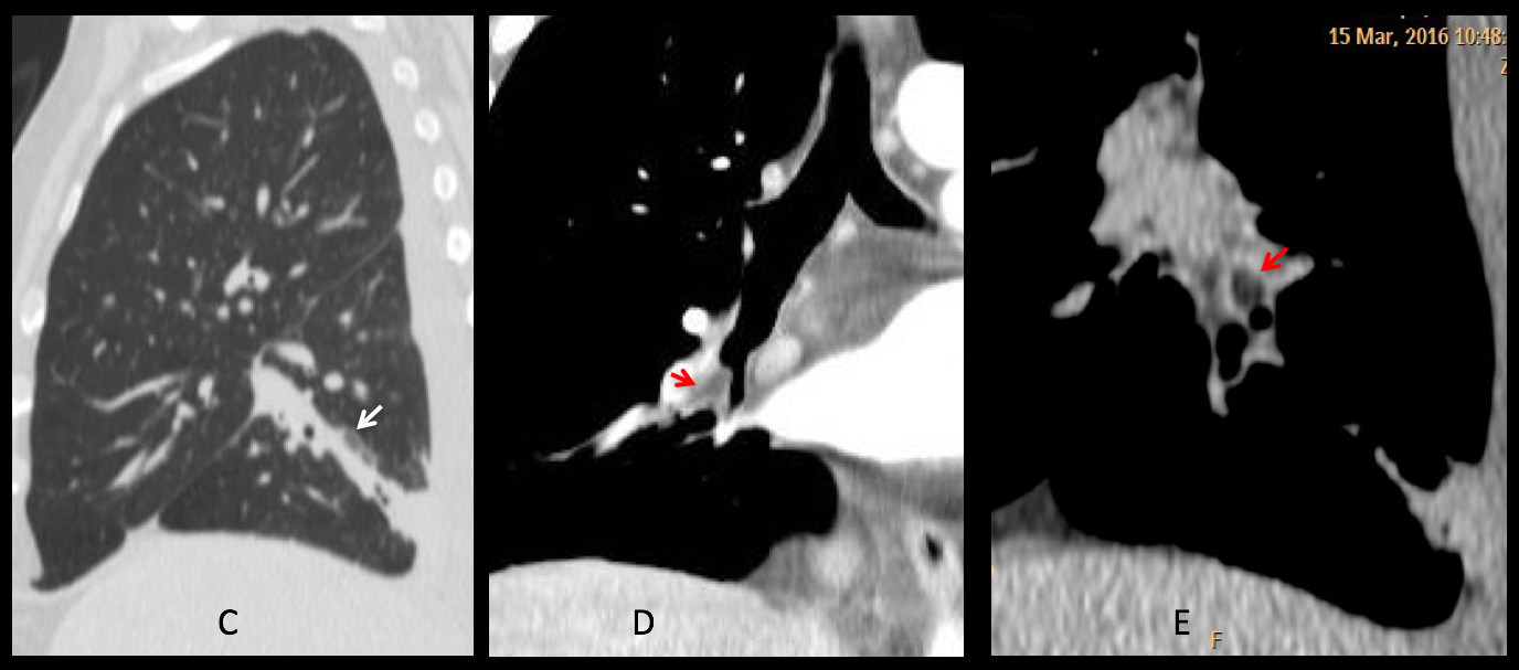
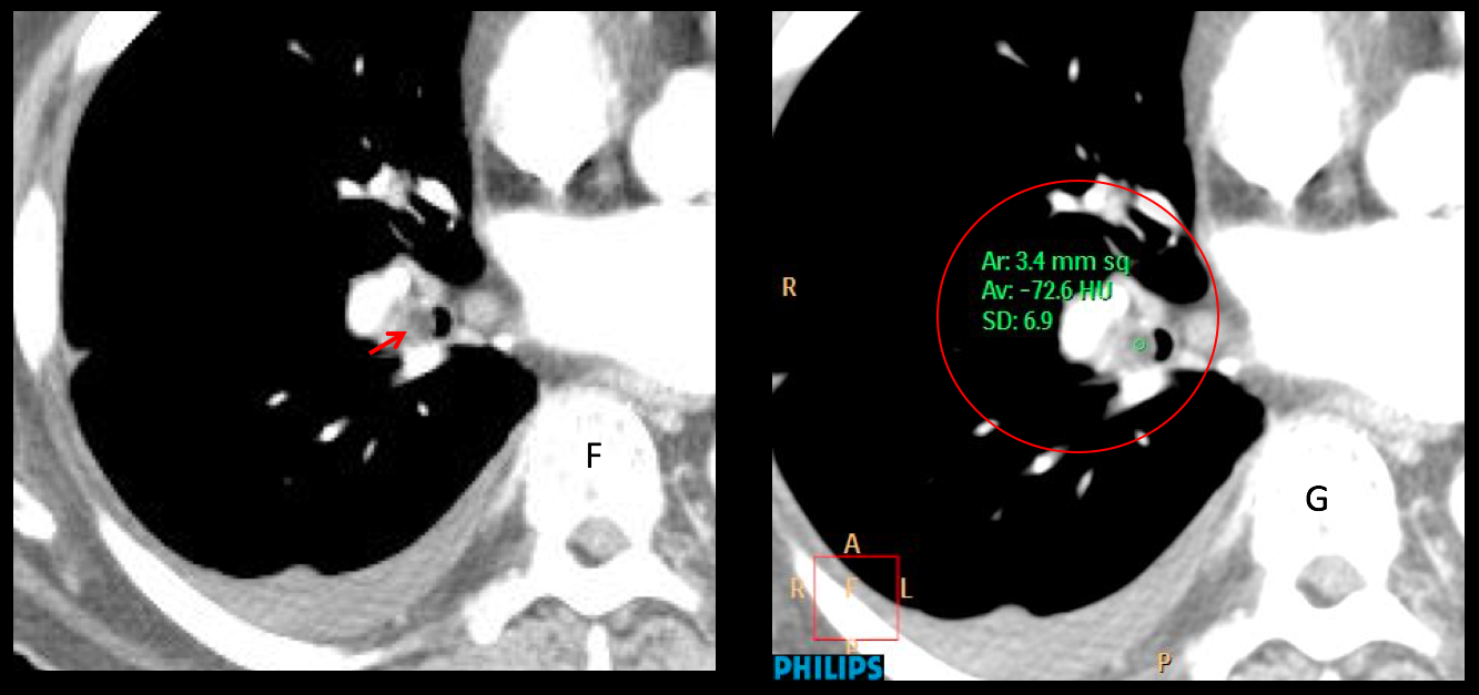
Enhanced axial CT confirms the endobronchial lesion (F, red arrow), which measures -72 H.U. (G, circle). Endobronchial lipoma was suspected and confirmed at bronchoscopy.
Final diagnosis: endobronchial lipoma of posterior segmental RLL bronchus
Congratulations to MK and MP who, with a little bit of help, made the correct diagnosis.
Endobronchial lipomas, also called lipomatous hamartomas are not infrequent in my experience and easily diagnosed by CT. As a bonus point, the last figure shows my first lipoma, seen in the eighties and discovered because of the descended right hilum.
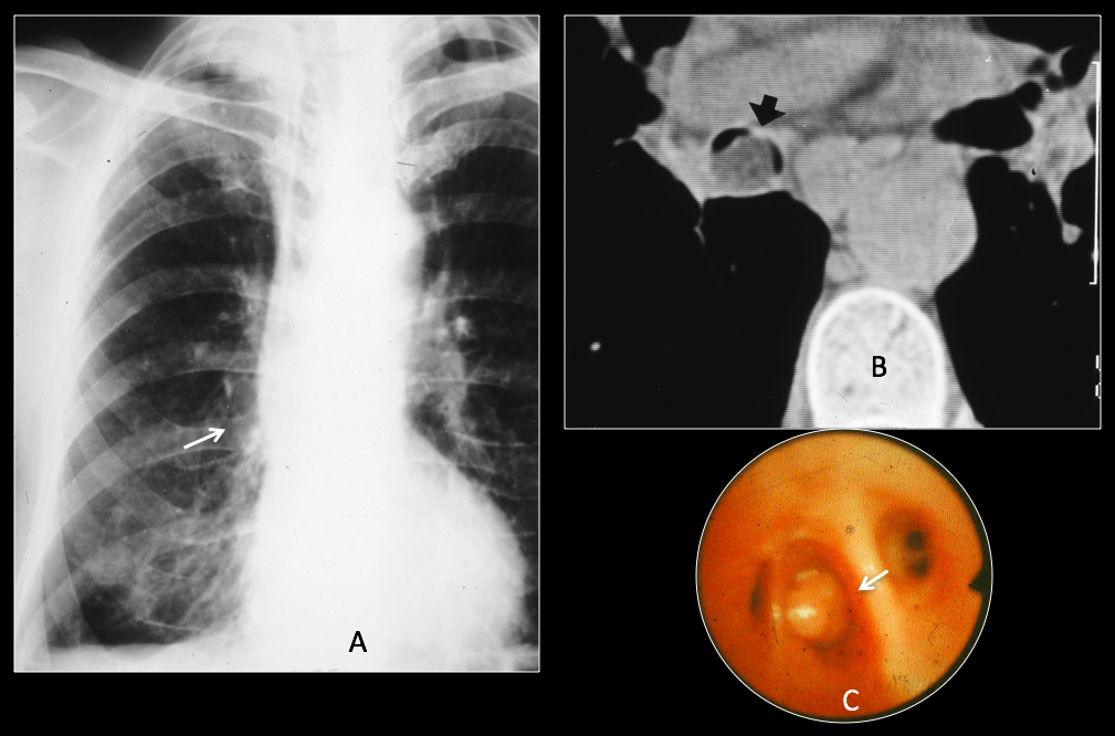
56-year-old male with chronic cough. The right hilum is displaced downward (A, arrow). Unenhanced axial CT shows a low-density mass in the intermediary bronchus (B, arrow). Lipoma confirmed at bronchoscopy (C, arrow).
Teaching point: in lobar or segmental lesions of the lung always consider the possibility of an endobronchial lesion. It led to the diagnosis in this patient who came for an unrelated process.