Dear Friends,
Happy New Year! To start 2018 with an easy case, I am showing pre-op PA radiograph and CT of a 49-year-old woman with carcinoma of the breast.
Diagnosis:
1. Metastasis
2. Neurogenic tumour
3. Duplication cyst
4. Any of the above
Check the images below, leave your thoughts in the comments section and come back on Friday for the answer.


Click here for the answer
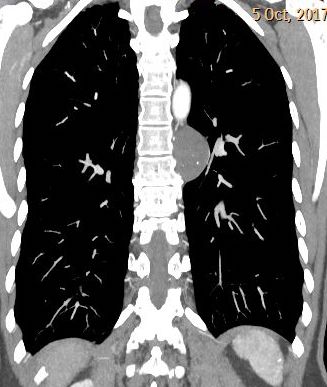
Findings: the PA chest radiograph shows a left mediastinal mass (A, arrow). Coronal CT confirms a posterior low-density mass with punctiform calcification (B, arrow).

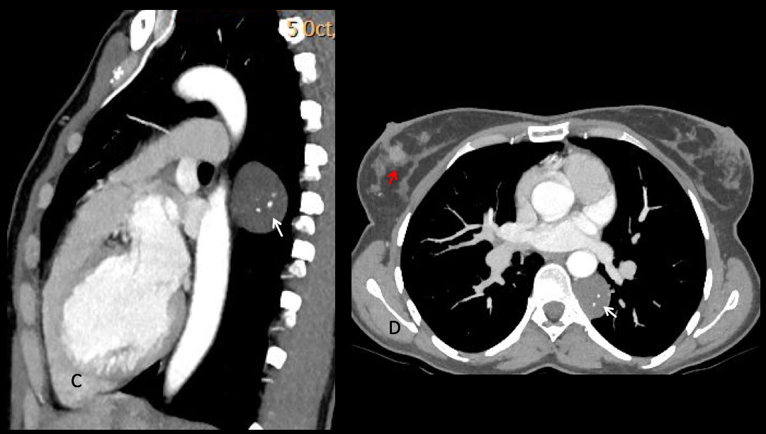
Axial and sagittal views show several calcifications within the mass (C-D, arrows). The location, low density and the presence of calcium strongly suggest
a neurogenic tumour. An additional finding is the depiction of the tumour in the right breast (D, red arrow).

Final diagnosis: unsuspected neurogenic tumour in a patient with carcinoma of the breast.
Most of you did very well and suggested the right diagnosis. Special mention to Yulia and Mauro, who were the first.
Teaching point: calcium within a low-density mediastinal mass excludes a cystic lesion. In this case the posterior location favored a neurogenic tumour.


Neurogenic tumor
Hello!
There is a rounded and mildly heterogeneous rounded mass in the left posterior mediastinum, with punctate calcifications. I can also see discrete vertebral scalloping. My first hypothesis is neurogenic tumor (number 2).
Quiste neurogenico
Even I think it could be a neurogenic tumor
Neurogenic tumour
Extramedullary Hematopoiesis
Neurogenic tumour
2. Neurogenic tumour
Heterogeneous rounded mass in the left posterior mediastinum with vertebral scalloping probably neurogenic tumor