
Dear Friends,
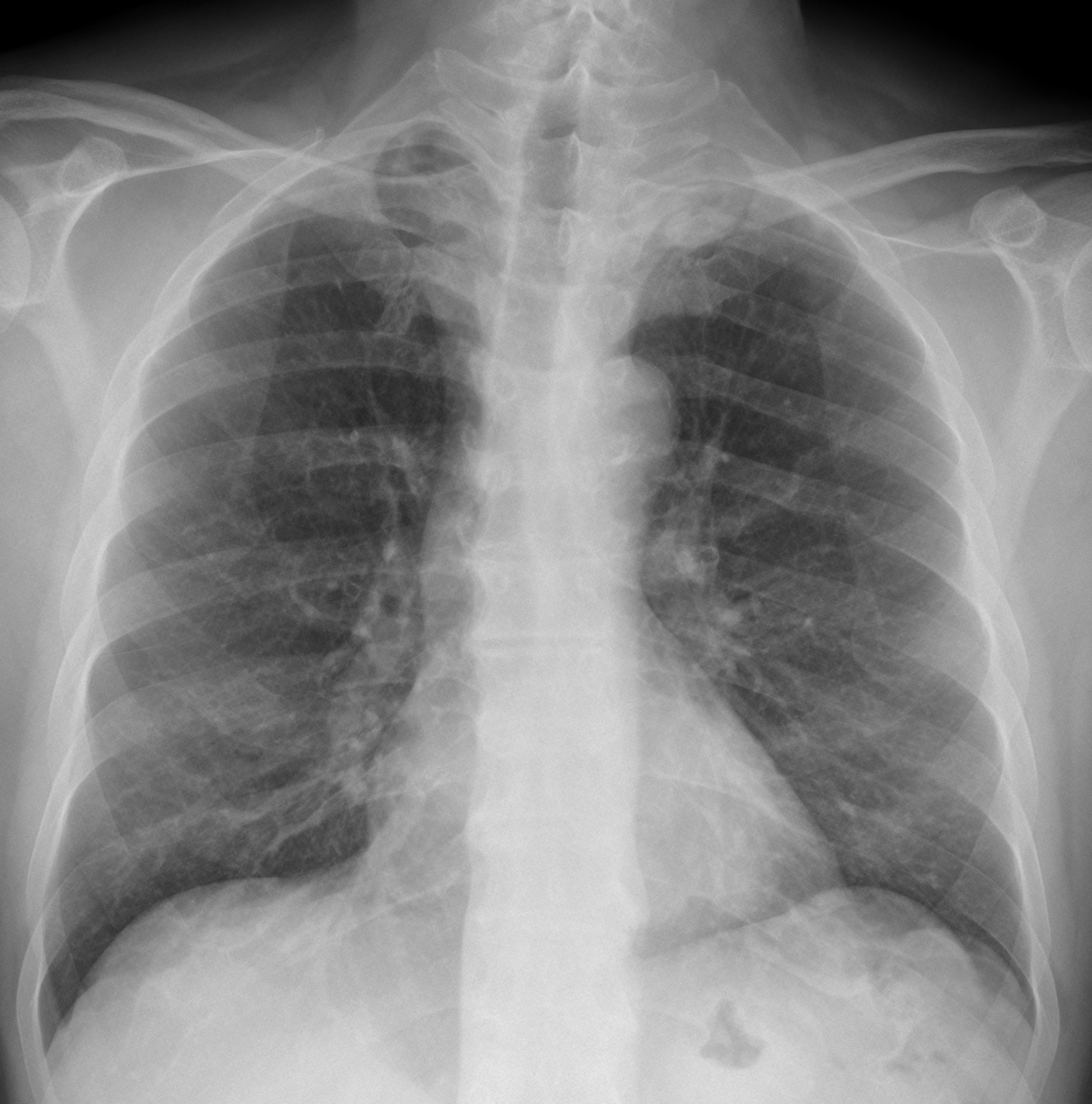
Today, I am showing radiographs of a 68-year-old man with pain in the chest. What do you see?
Check the images below, leave your thoughts in the comments section, and come back on Friday for the answer.
Click here for the answer
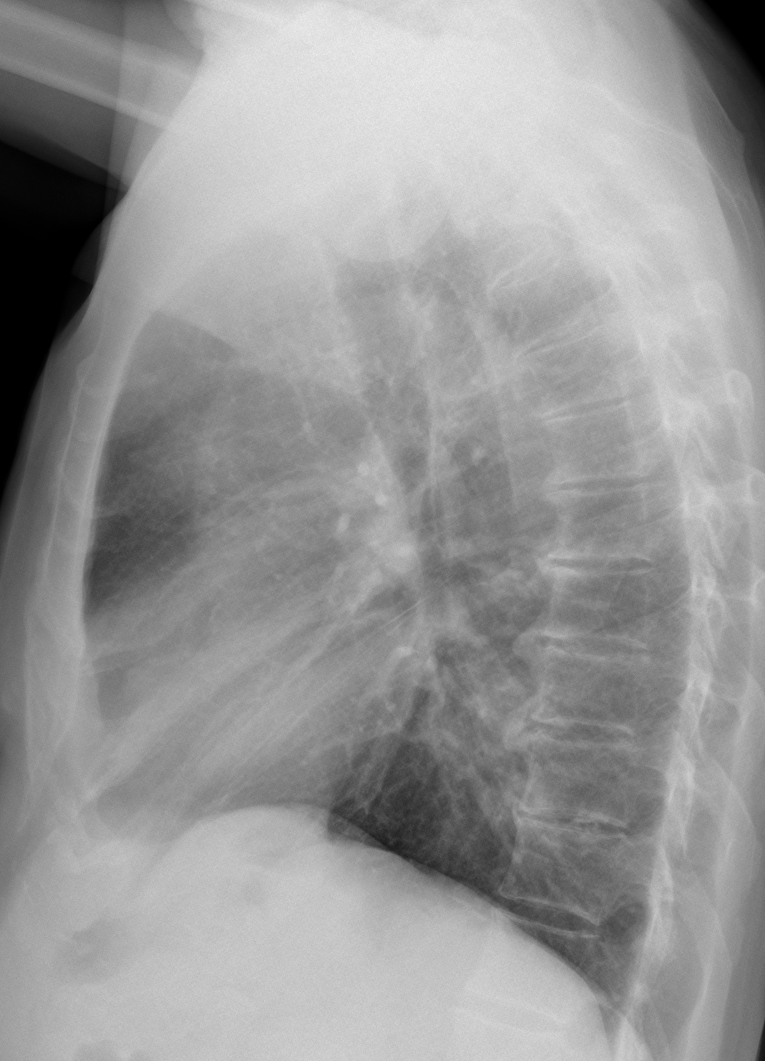
The main finding is a rounded left apical opacity visible only in the PA radiograph (A-B, arrows). The lateral view is unremarkable.
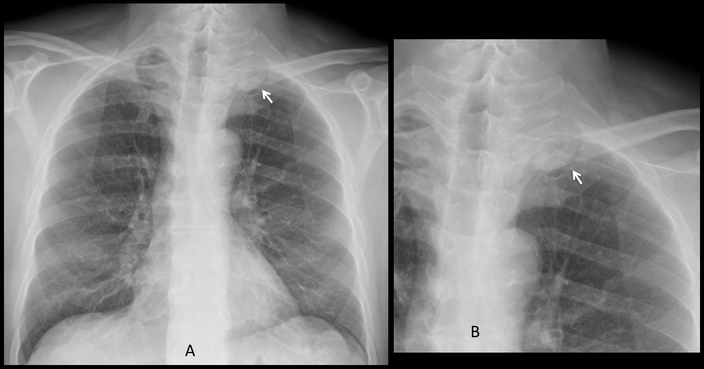
The patient complained of pain in the left shoulder. Coronal and axial enhanced CT conforms the apical mass (C-D, arrows). Needle biopsy came back as adenocarcinoma.
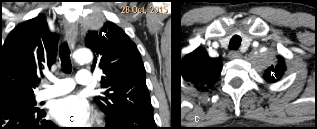
Final diagnosis: Pancoast tumour
Congratulations to the three initial brave correspondents (MK, Coffee and Amago) who made the diagnosis.
Teaching point: the pulmonary apex is a dark area in the chest radiograph and tumours in this location are missed often. Don’t forget to look at the apices in the PA view.


