As promised, here are the two additional images. Do they help you to reach a conclusion?
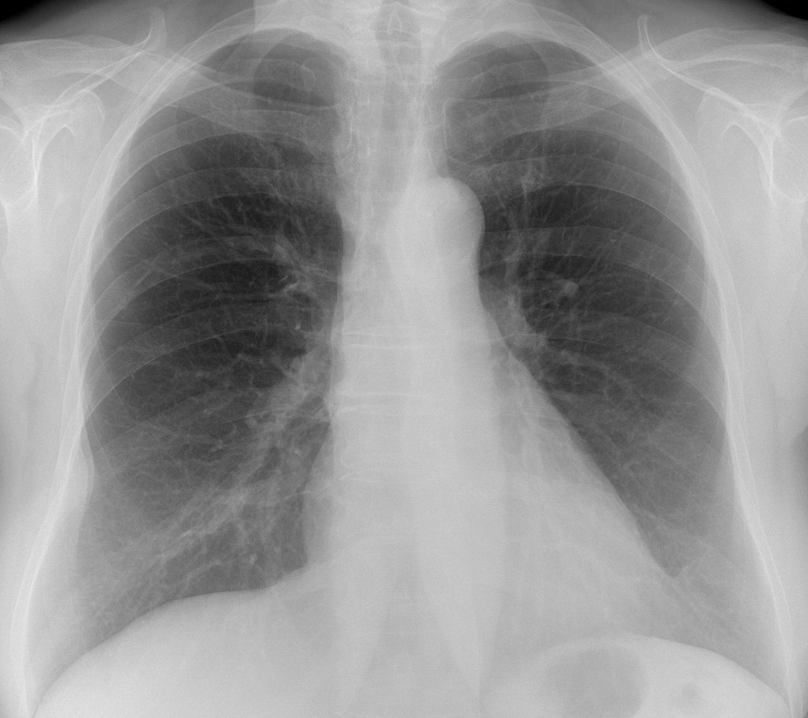
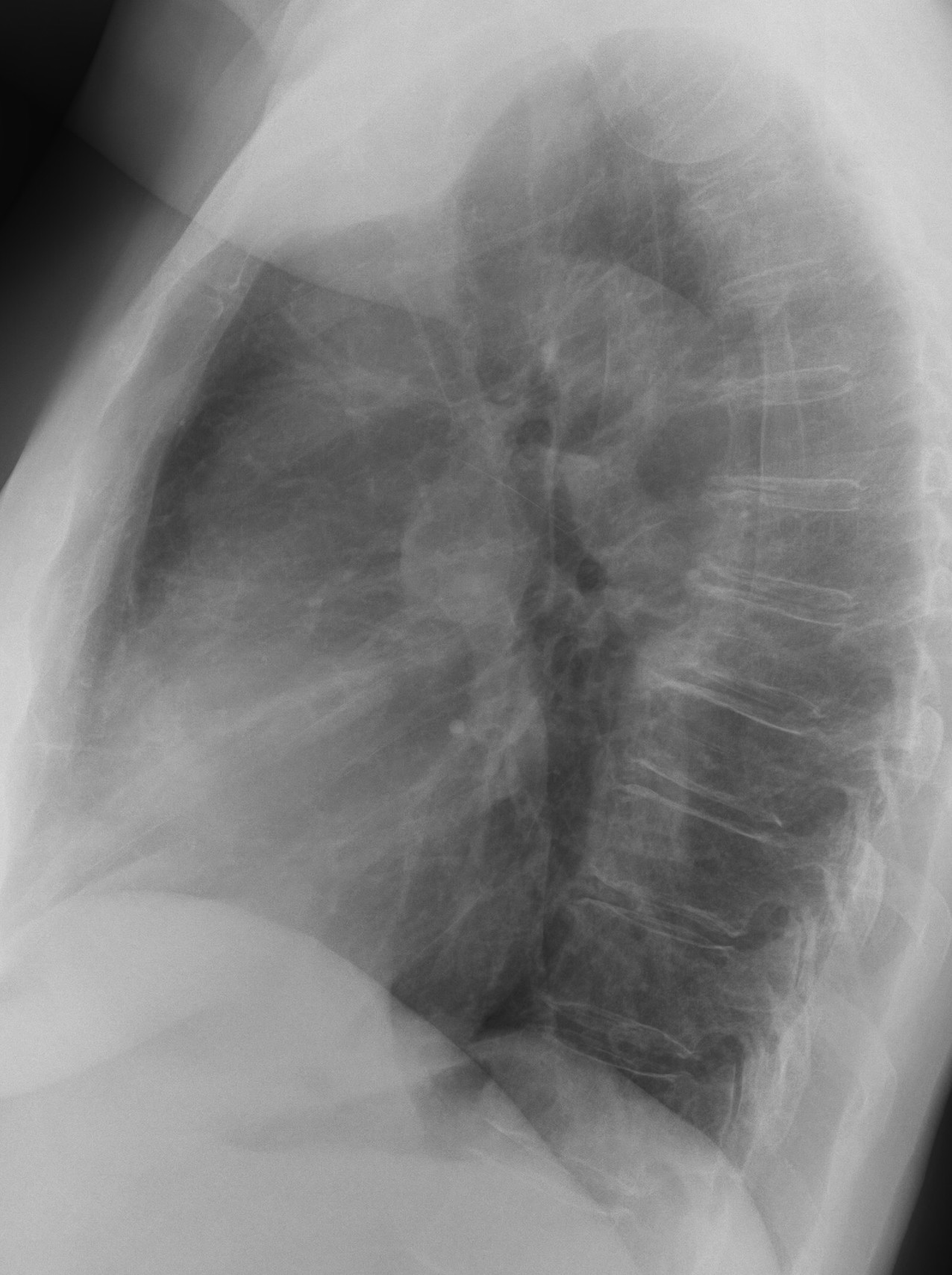
Findings: the PA radiograph shows mild compression of the right wall of the cervical trachea due to a known goiter. The main finding is the presence of an extrapulmonary lesion in the lower right hemithorax (A, arrow) with possible enlargement of the underlying rib giving a double contour to the lesion (B, arrow).

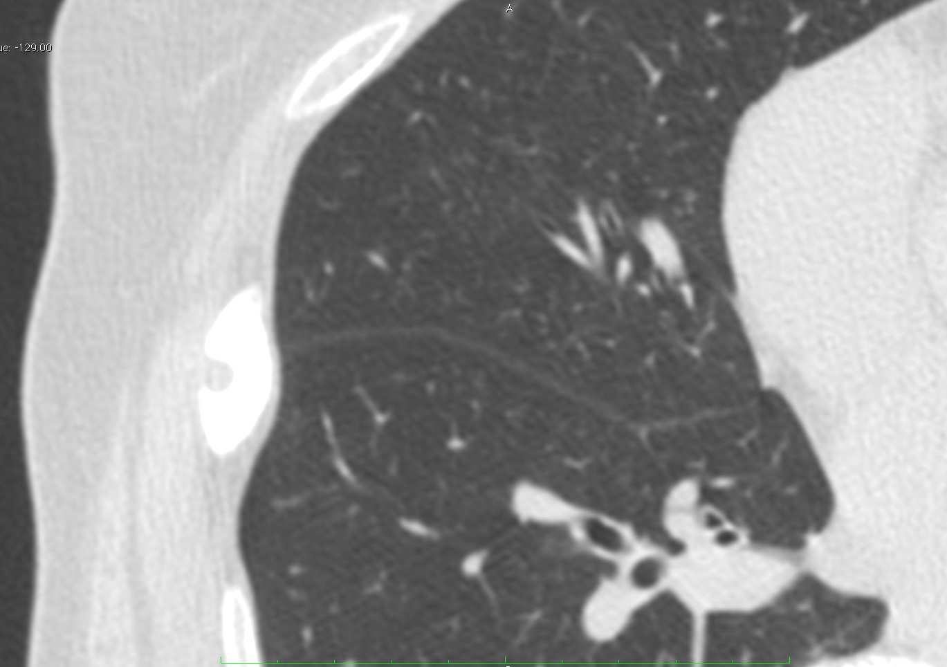
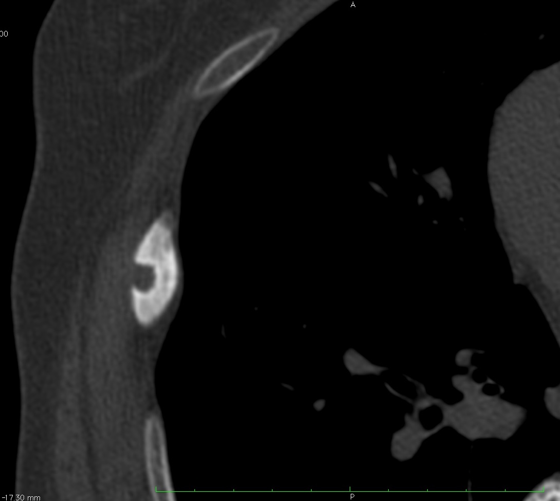
Unenhanced axial CT shows a widened sclerotic rib with a well-defined punched-out lesion in the outer cortex (C-D, arrows) with sclerotic border.
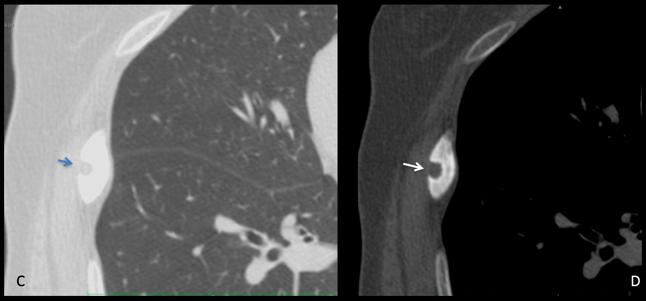
As I mentioned when presenting the case, I don’t have a definitive diagnosis. The lesion is clearly slow growing, as evidenced by the sclerotic border. Certainly it is not an osteoid osteoma (no pain and no swelling of soft tissues). The peripheral location and the aspect go against enchondroma. I cannot rule out fibrous dysplasia, a common lesion in the rib, but it does not look like one to me.
After interrogation, the patient recalled a previous fall while in the country many years ago, hurting her right side. My explanation is that she got a sharp vegetal foreign body in the rib at that time, which ended in a foreign body rib granuloma.
The patient is asymptomatic and I will do a CT follow-up in six months; will share the results with you here on the ESR blog.
Tentative diagnosis: Foreign body granuloma of the rib (unproven)
It is difficult to congratulate anyone, since I don’t have the right answer. In my opinion, Sadaf and Coffee were the people who better analysed the findings.