Dear Friends,
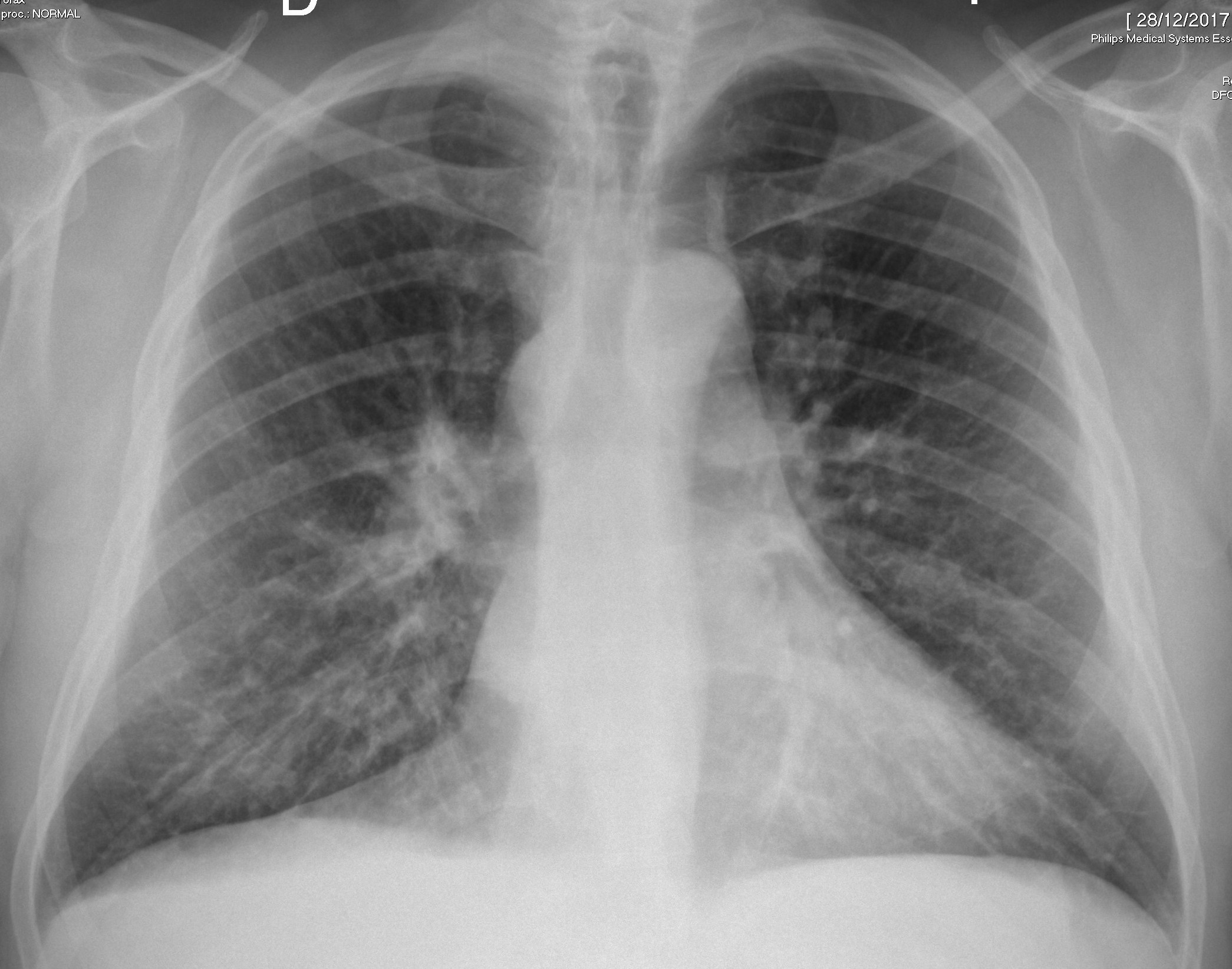
I want to present today a case that I saw two months ago. The radiographs belong to an asymptomatic 55-year-old man. Will show more images on Wednesday.
Check the images below, leave your thoughts in the comments section, and come back for the answer on Friday.
Diagnosis:
1. Enlarged azygos vein
2. Enlarged ymph node
3. Mediastinal mass
4. Any of the above
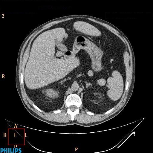
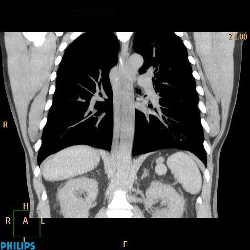
As promised, here are two additional CT images. What do you see?
Click here for the answer
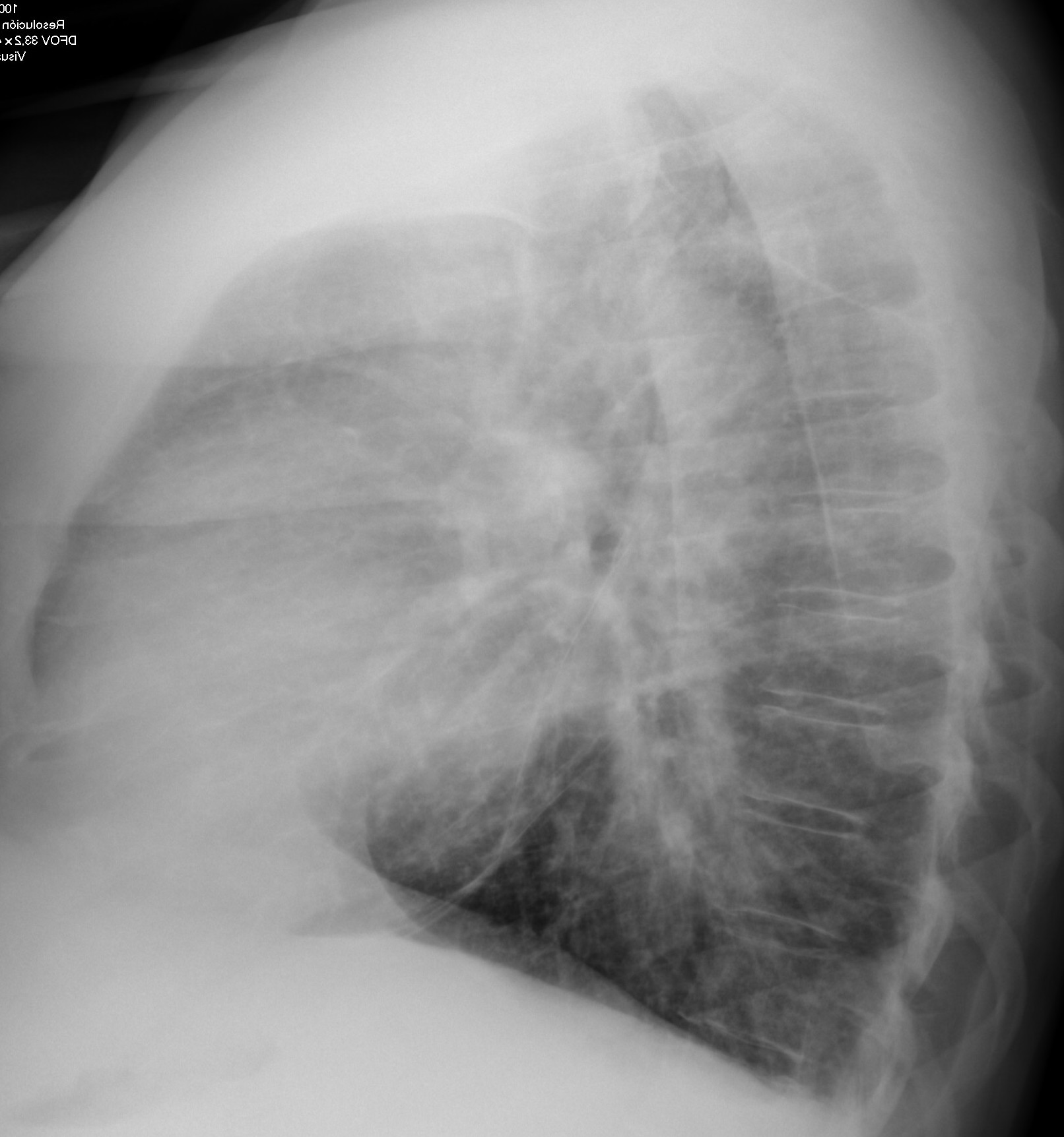
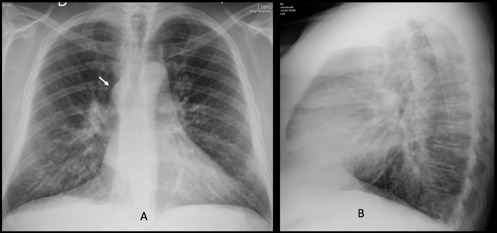
Findings: the PA radiograph shows a mediastinal mass at the level of the azygos vein (A, arrow), similar to the one seen in
case 178. The same differential diagnosis applies. The lateral view (B) is unremarkable.
Unenhanced coronal and axial CT show an enlarged azygos vein (C-D, arrows). The inferior vena cava is absent. There are two nodules in the left upper quadrant of the abdomen (B-C, red arrows).
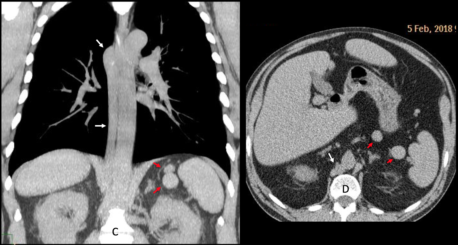
The findings are characteristic of a partially absent IVC which drains through the azygos vein. The nodules in the left upper quadrant represent accessory spleens.
I am showing this case to complement case 178, which had similar findings in the plain films that were not documented by CT. Also, both cases have been seen in a short period of time.
Final diagnosis: azygos continuation of IVC with polysplenia
Congratulations to MK, who was the first to suggest the diagnosis
Teaching point: congenital anomalies are not unusual. It is important to recognise them to avoid confusion with more serious conditions.


