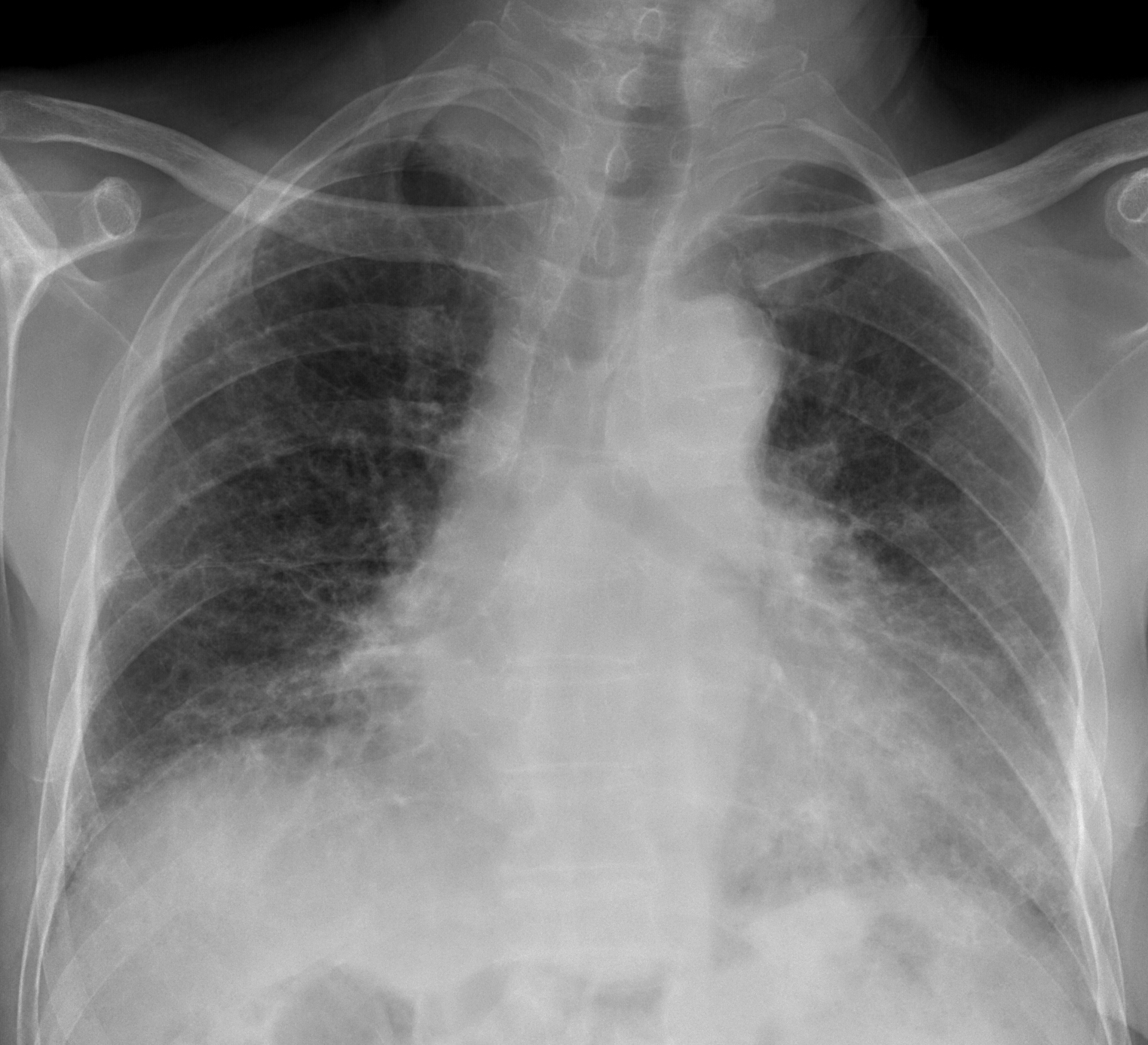
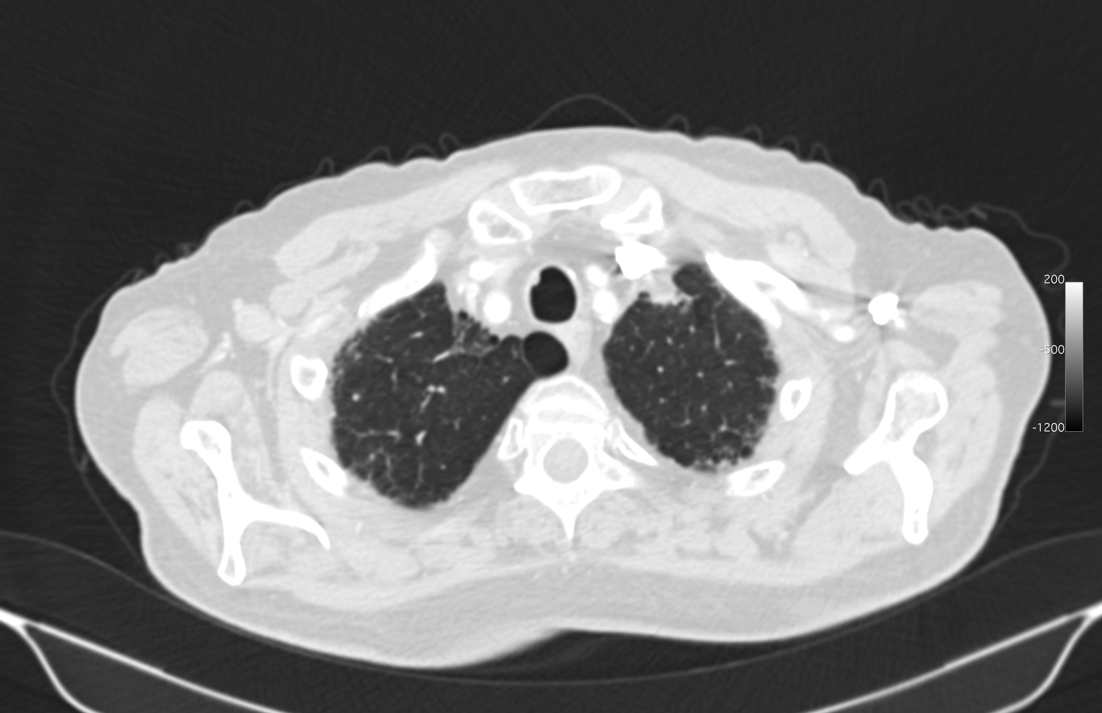
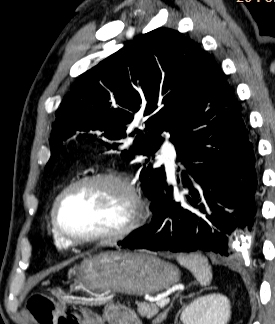
This week’s radiographs belong to a 68-year-old man with pulmonary fibrosis and dyspnoea. Check the images below and leave your thoughts in the comments section. New images will be added on Wednesday, followed by the answer on Friday.
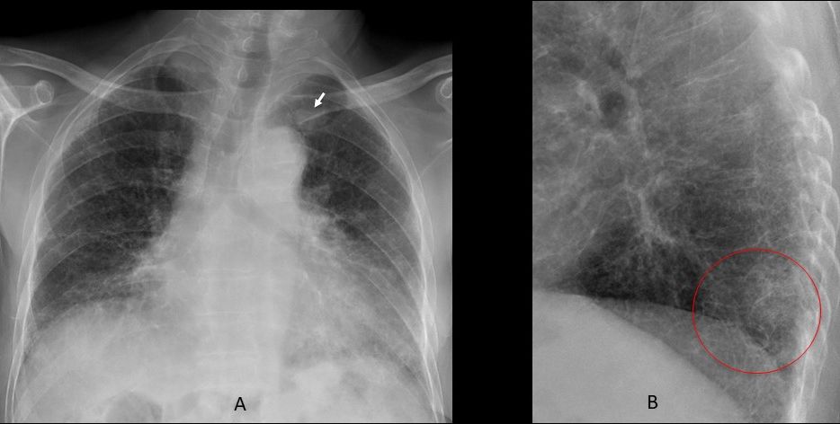
Findings: Aside from the extensive changes of interstitial disease, a left infraclavicular nodule is seen in the PA radiograph (A, arrow). The lateral view shows a peanut-like image in the posterior clear space (B, circle).
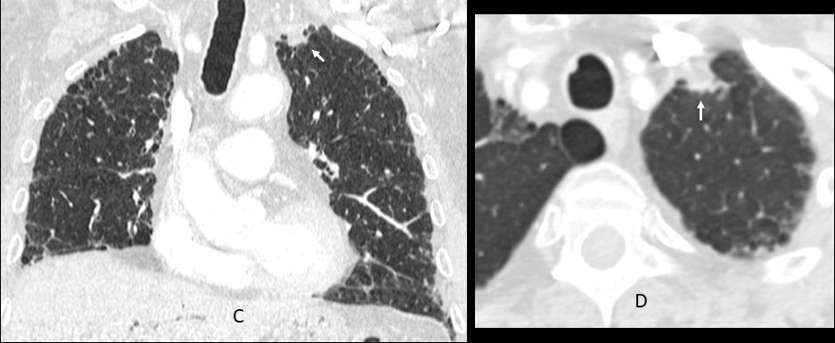
Coronal and axial CT confirms the presence of the apical lung nodule (C-D, arrows).
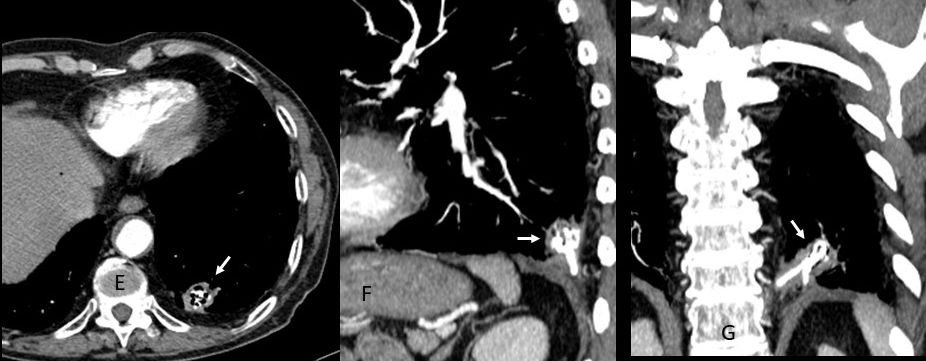
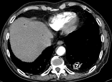
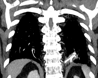
The most interesting finding is seen at the base of the left lung: the enhanced axial CT shows a cavity with calcified content (E, arrow), which looks like a candelabra in the sagittal reconstruction (F, arrow) and like a loop in the coronal CT (G, arrow). This appearance brings to mind calcified strands of fungi (hyphae).
The patent was operated on and a carcinoma of LUL was found. The lesion at the LLL represented a cavity filled with aspergillus fungi.
Final diagnosis: Carcinoma of LUL and calcified aspergilloma of LLL.
This is an unusual case and there were very few responses. Have to congratulate MK because she saw the apical nodule in the chest film. And also, Genchi Bari for his unrelenting enthusiasm (and his support for Bari FC!)