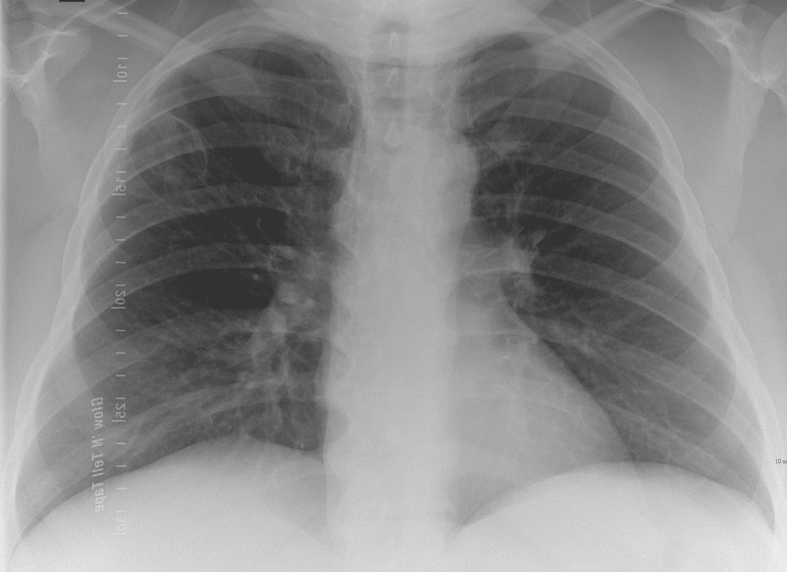
Today I am presenting a recent case, seen last January. Below is a preoperative PA chest radiograph for bariatric surgery in a 44-year-old woman. Check the image and leave me your thoughts in the comments section. I will show more images on Wednesday, followed by the final answer on Friday.
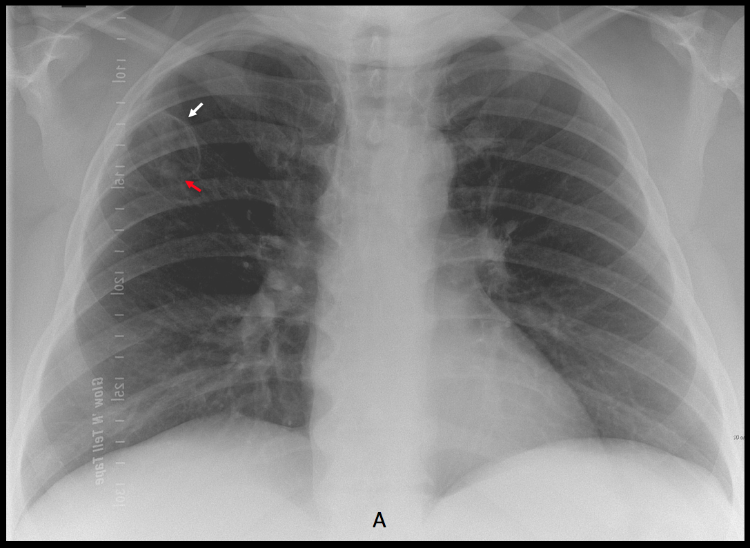
Findings: the preoperative PA chest radiograph shows a thin-walled cavity in the RUL (A, arrow) with a small nodule inside (A, red arrow).
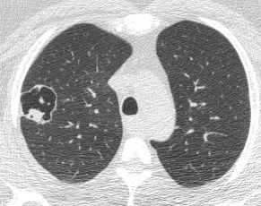
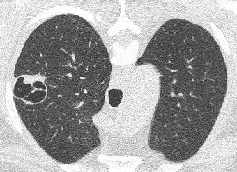
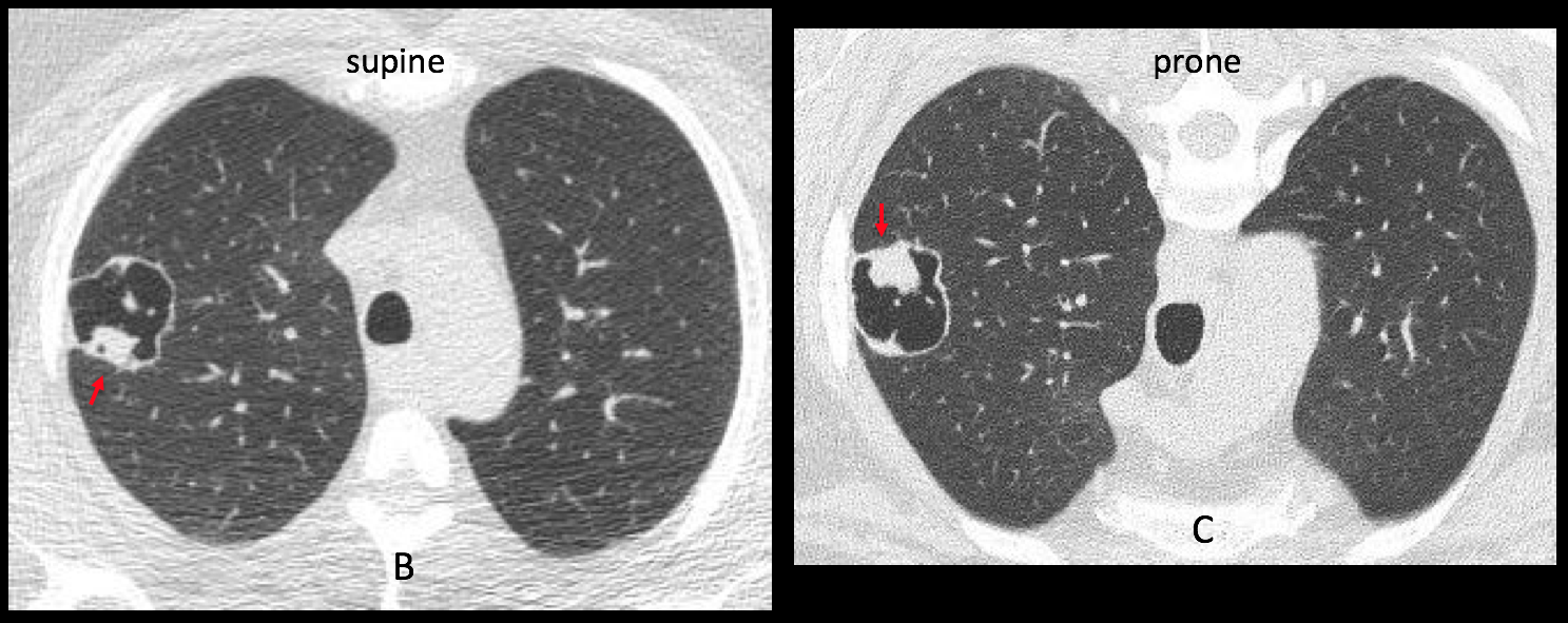
Axial CT confirms the thin-walled cavity and the nodule, which does not move in the prone position (B-C, red arrows).
An additional finding is the presence of a pulmonary infiltrate in the proximal RUL bronchus (D-E, arrows).
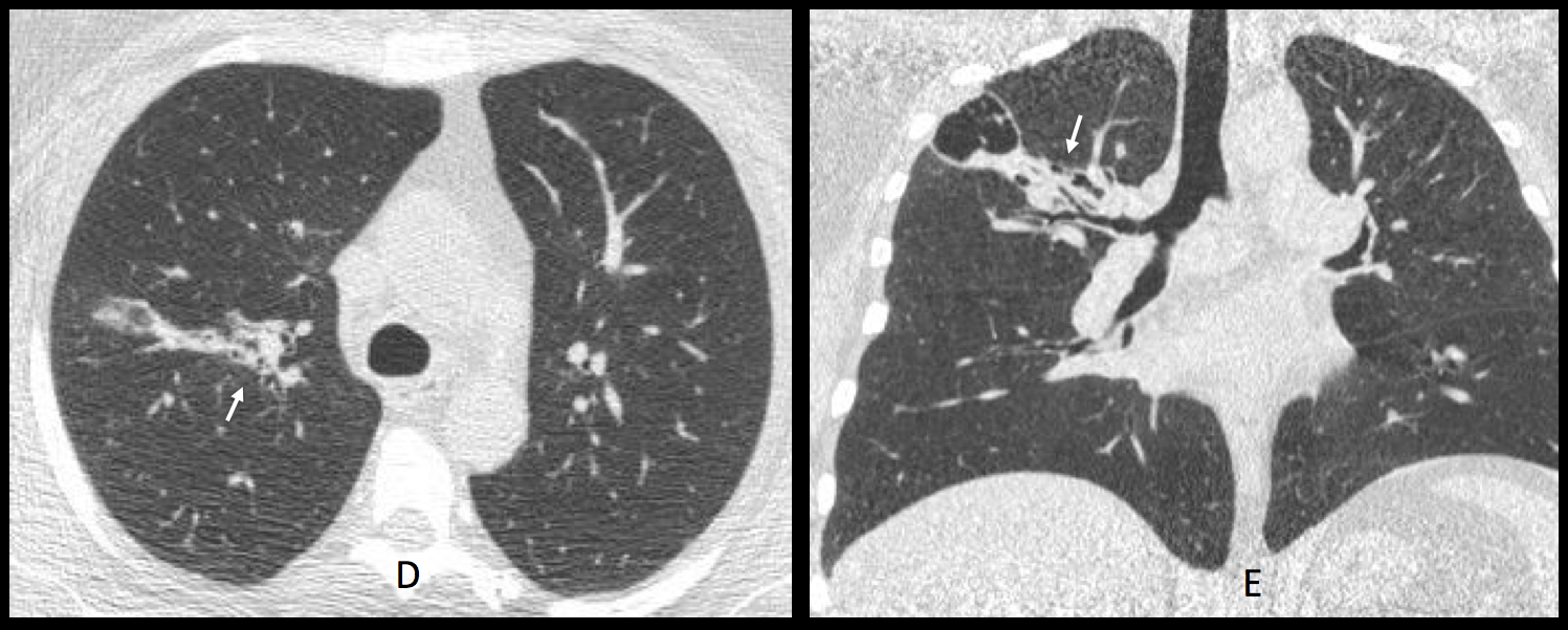
This case presents conflicting evidence: on one side, the nodule does not move, suggesting an intracavitary neoplasia. On the other hand, the thin walls of the cavity point to a benign lesion and the pulmonary infiltrate suggests an inflammatory process. In my opinion, this last finding is the one that carries more weight towards benignancy. Bronchoscopy was done and demonstrated Aspergillus in the proximal bronchi.
Diagnosis: Aspergillus infection, probably a mild form of semi-invasive aspergillosis.
This was a difficult case and I want to congratulate all the participants because all of you did your best.
Teaching point: when confronted with a case with conflicting finding, choose the one that, in your experience, is the most likely to be of help.