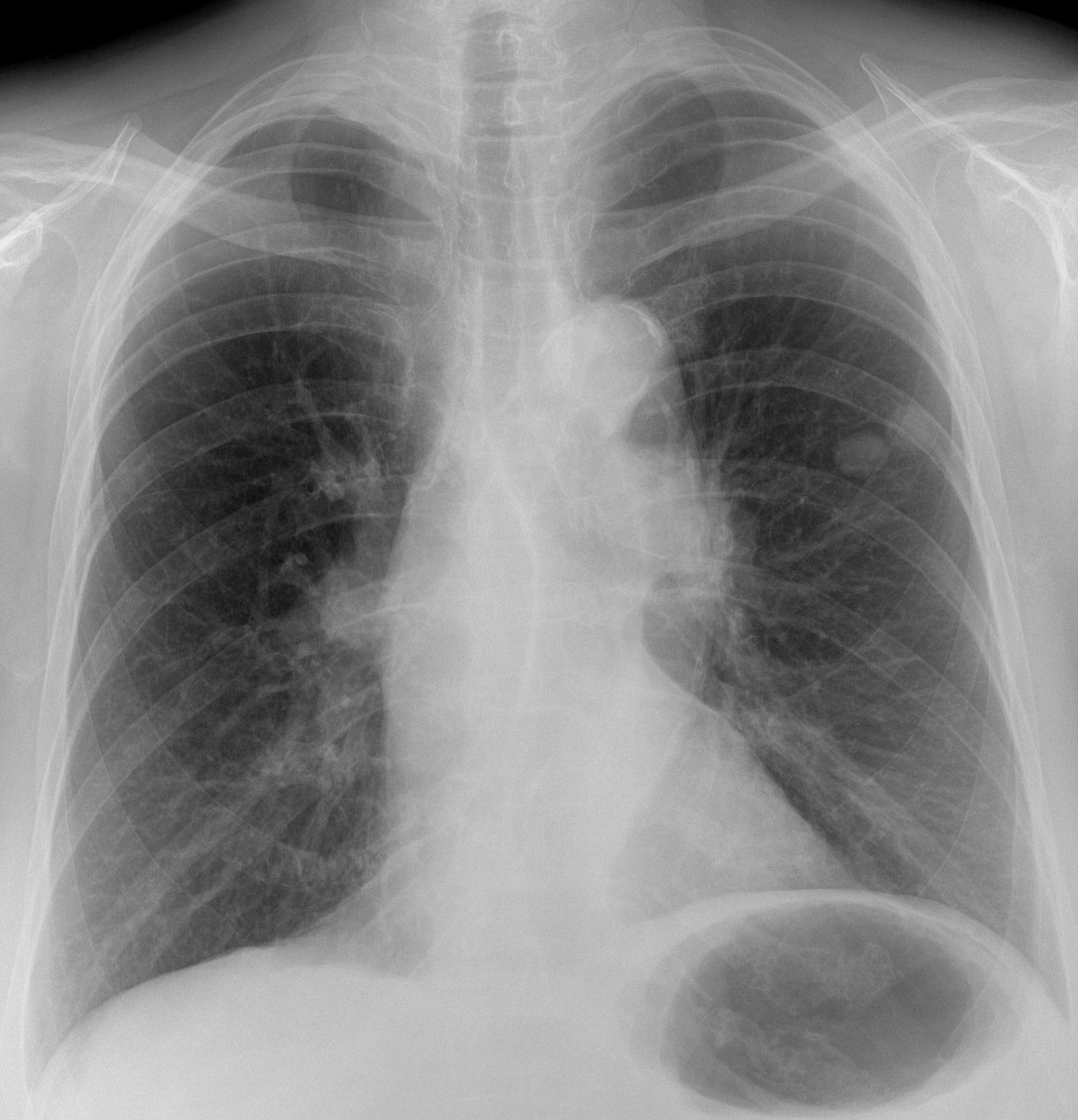
Today I am showing a pre-op chest radiograph for varices of a 79-year-old woman. A radiograph taken five years ago was normal.
What do you see?
Check the image below and leave your thoughts in the comments section. Will show more images on Wednesday and the answer on Friday.
Findings: PA radiograph shows two nodules in the left middle lung field (A, arrow). In addition, there is a mass in the left hilum (A, red arrow). These findings were not present in a previous film taken five years earlier (B).

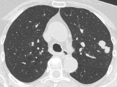
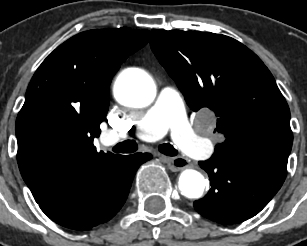
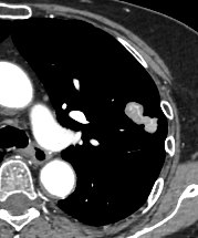
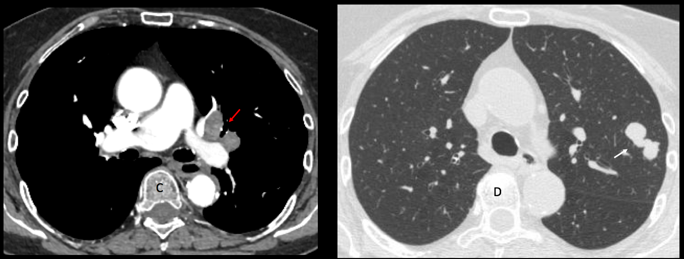
Enhanced axial CT shows lymph nodes in the left hilum (C, red arrows), as well as a dumbbell-shaped lesion in the left lung (D, arrow).
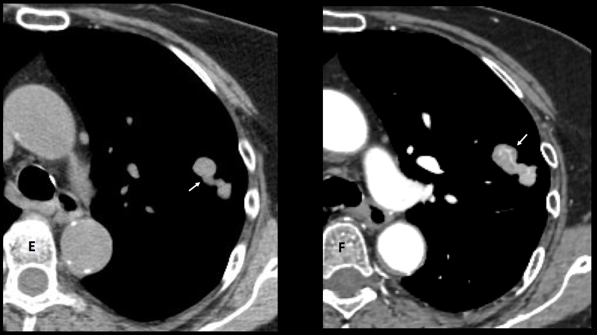
The lesion has meandering vessels within (E-F, arrows) and enhances 35 H.U. after contrast. A needle biopsy returned as small-cell carcinoma.
Final diagnosis: small-cell carcinoma of the lung with hilar metastases.
Congratulations to Trinity, who made an accurate diagnosis.
Teaching point: this case puzzled me. My original diagnosis was malignant fibrous tumour of the lung because of the odd shape and the presence of arterial vessels inside.
Unfortunately, reality defeated me. The lesson could be: always think of a rare manifestation of a common disease rather than a common manifestation of a rare disease. I stand corrected.