Today we are presenting a pre-op chest radiograph for knee surgery of a 48-year-old woman. What do you see?
Check the image below and leave your thoughts in the comments section. More images will be shown on Wednesday, and the final answer on Friday.
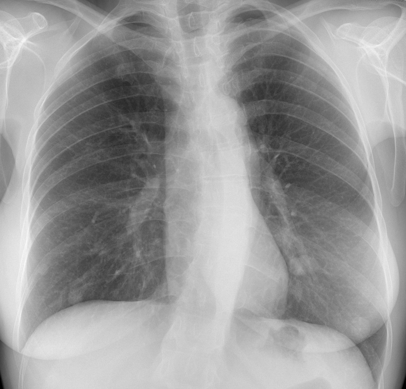
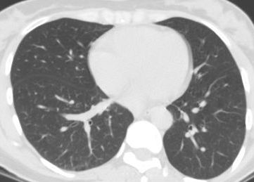
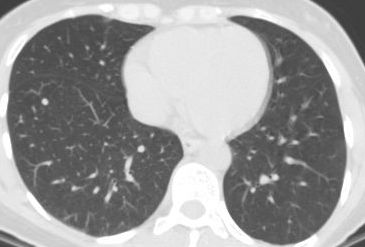
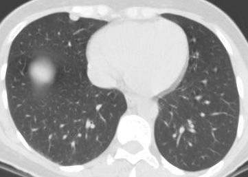
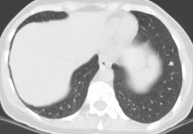
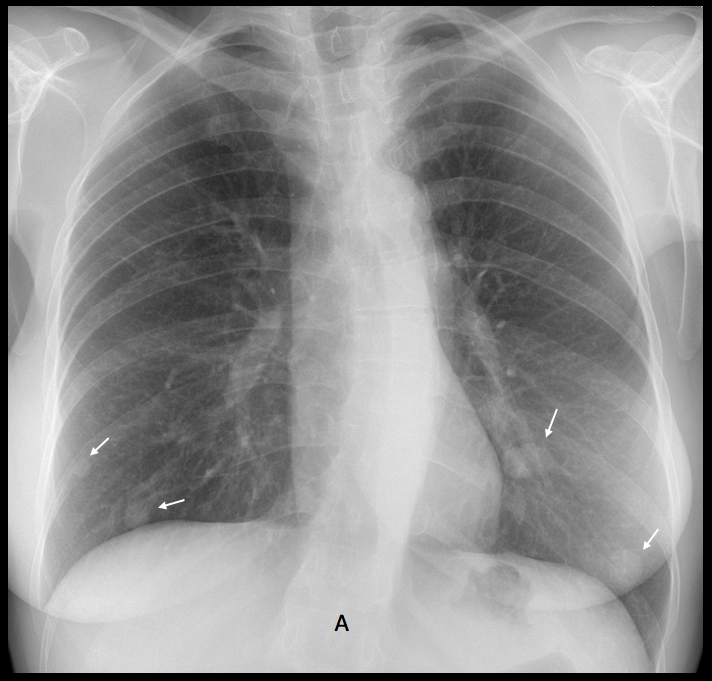
Findings: PA chest shows several nodules of unequal size in both lung bases
(A, arrows).
CT taken five years earlier for a study of hypertension demonstrates the same nodules, albeit smaller (B-E, arrows).

The differential diagnosis of slow-growing lung nodules is ample. The location in the lower lungs suggests haematogenous seeding and, in a female patient, metastasising leiomyoma should be considered. This particular patient had a history of uterine leiomyomas resected. At the time of the CT, a 7mm nodule in the RML was resected and diagnosed as leiomyoma.
At the present time the patient (a physician) refuses a new CT and/or needle biopsy.
Final diagnosis: Benign metastasising leiomyoma of lung.
Congratulations to all of you who faced a difficult case with dignity.
Kudos to Marco S, who suggested the correct diagnosis.
Teaching point: clinical history, location of the disease and previous studies are of great help in suggesting the right diagnosis.