Today’s radiographs belong to a 68-year-old man in whom a chest abnormality was found in a routine check-up.
Check the images below, leave your thoughts in the comments section, and come back for the answer on Friday.
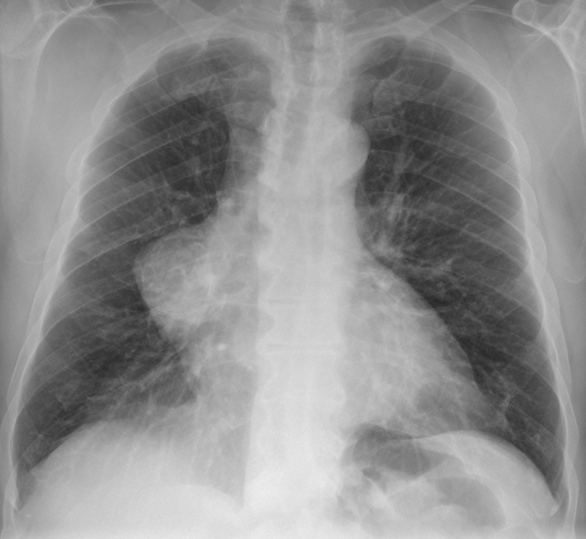
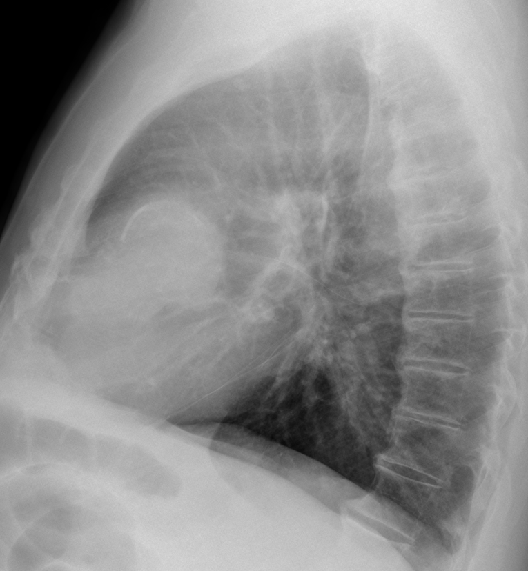
Findings: chest radiographs show an obvious anterior middle mediastinal mass (A-B, arrows) with thick peripheral curvilinear calcification, better seen in the lateral view (B, red arrow).

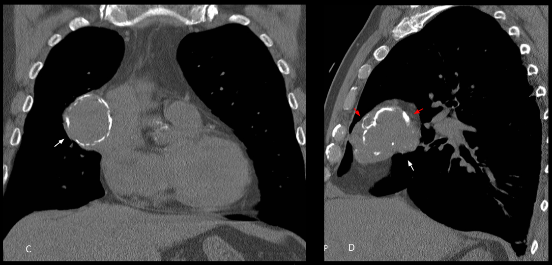
Unenhanced coronal and sagittal CT confirms a solid mediastinal mass with coarse irregular peripheral calcification (C-D, arrows). The irregularity and coarseness of the calcium is better seen in the sagittal reconstruction (D, red arrows).
Diagnosis after surgery was Type I thymoma.
Chest radiographs were not easy to interpret, and I congratulate all of you for trying. In my opinion, the calcium in plain film was too thick to think of a vascular or cystic lesion, leaving open the option for a solid calcified lesion. In my absolutely biased opinion I wish to congratulate Adel as the winner.
Final diagnosis: Calcified thymoma
Teaching point: remember that thymomas may travel downwards and be found in the middle or lower anterior mediastinum.
Also remember that we have eradicated hydatid cyst in Spain!