Today I am presenting routine radiographs of a 77-year-old man with previous history of carcinoma of the bladder ten years ago. What do you see?
Check the images below, leave your thoughts in the comments section, and come back on Friday for the answer.
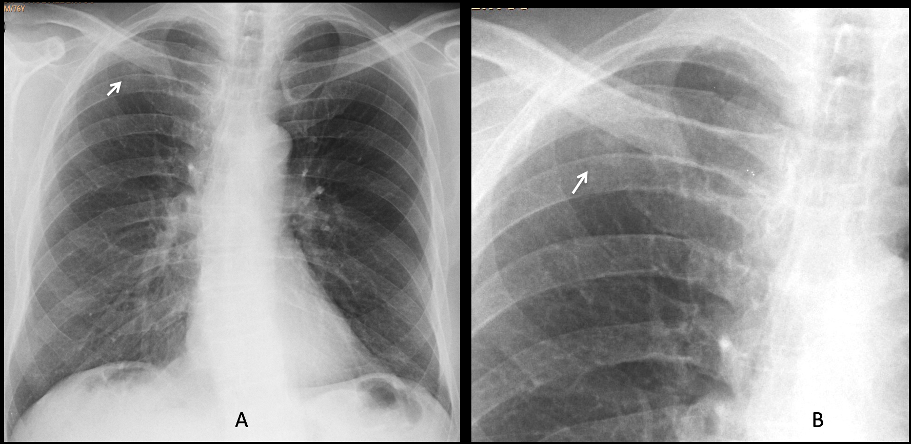
Findings: the PA chest radiograph shows a small right infraclavicular nodule (A-B, arrows), that was overlooked.
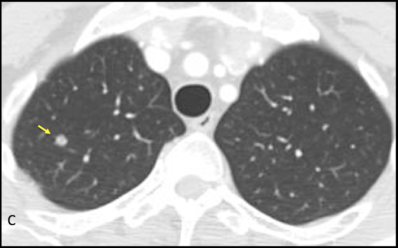
Interestingly enough, a CT taken six months earlier shows the nodule, smaller in size, that was also overlooked (C, arrow).
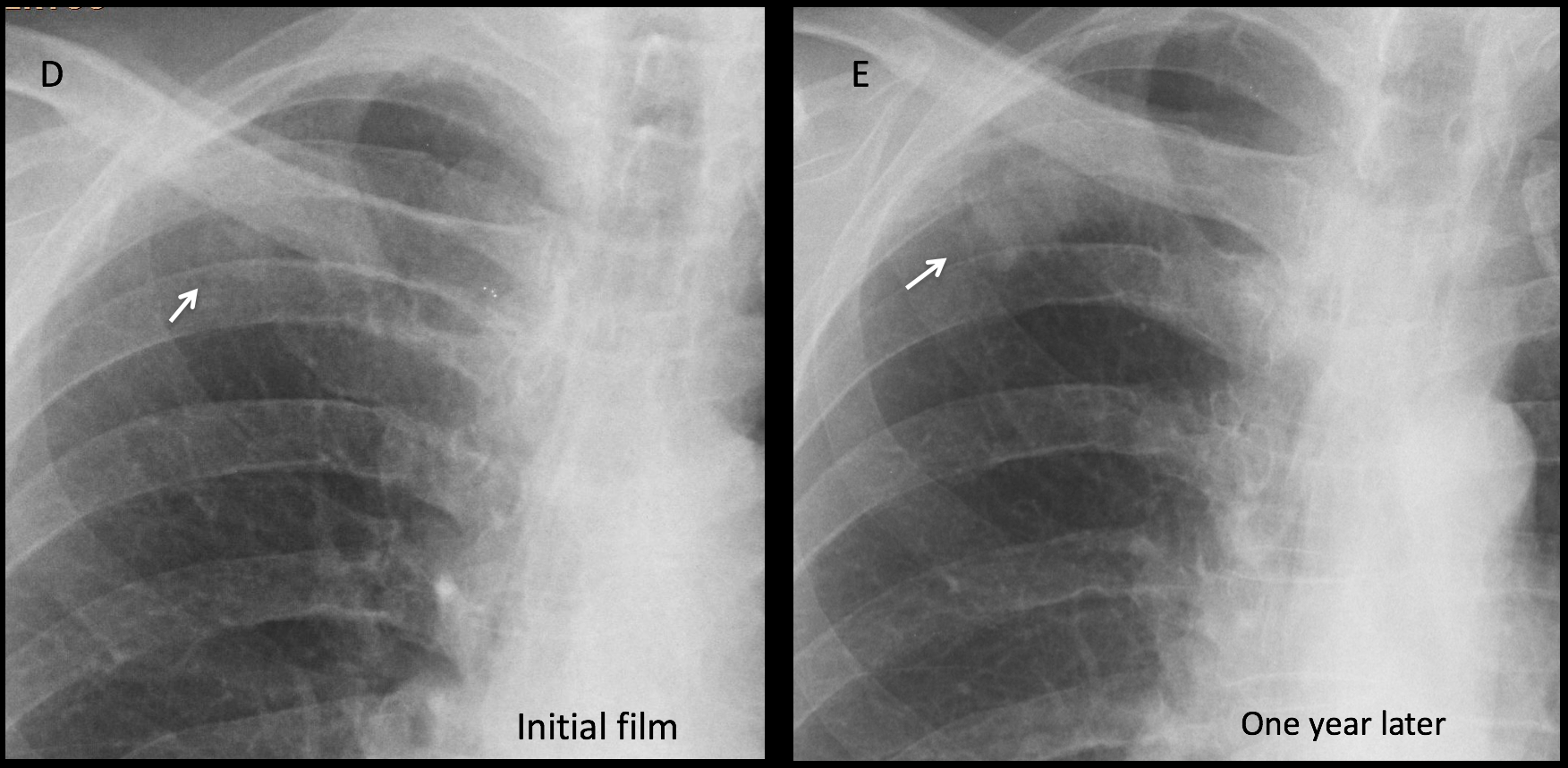
One year later the nodule has increased markedly in size (D-E, arrows) and this time it was noticed.
Considering the clinical history, the most likely diagnosis is carcinoma of the lung, which is frequently associated to carcinoma of the bladder. A metastasis is less likely, given the time interval since the original bladder surgery. The patient was operated on and a malignant melanoma was found.
Final diagnosis: melanoma of the lung.
Congratulations to Katarzyna who was the first one to see the nodule.
Teaching point: up to 50% of apical pulmonary nodules are missed in the chest radiograph. Remember to look carefully at the apices in every examination because many of them are malignant.


