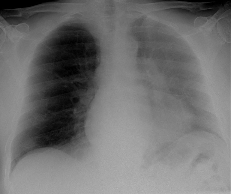
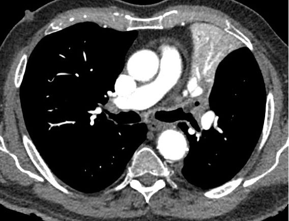
Hope you had a very good summer. I am back with a case from my good friend Alberto Villanueva. This PA chest radiograph belongs to a 57-year-old man with dyspnoea. More images will be shown on Wednesday.
showing enhanced axial CT.
Findings: the PA chest radiograph shows partial opacification of the left hemithorax. The left hilum seems increased in size (A, arrow) and there is blurring of the left heart border. In addition, there is increased lucency around the aortic knob (A, circle). All these signs are typical of LUL collapse in the PA film.

Enhanced axial CT confirms the LUL collapse (B, arrow). A low-density mass (-60 HU) is seen at the origin of the LUL bronchus (B-C, red arrows). This finding prompted me to make the diagnosis of endobronchial hamartoma/lipoma. At bronchoscopy, a peanut was found obstructing the LUL bronchus.
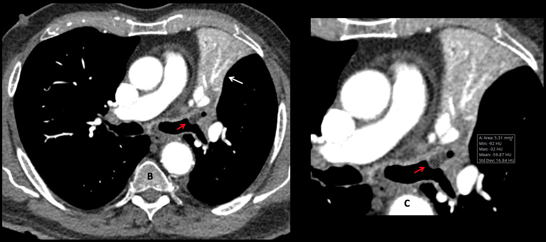
Final diagnosis: peanut obstructing the LUL bronchus and causing LUL collapse.
Congratulations to Alberto Montemayor, who was the first to diagnose LUL collapse.
I am showing this case because this is the first time that I have seen a fatty peanut in CT (Dr. Villanueva has shown me another case since). After learning this, I did a little experiment of my own, placing five different kinds of nuts in a plastic bag full of water and obtaining a CT. Two of them, macadamia nuts and peanuts floated to the top of the fluid and had fatty density. If any of you want to continue this nutty experiment, you have my blessing.
Teaching point: up to now, I considered any fatty endobronchial mass as a hamartoma/lipoma. From now on I have added peanuts to the differential diagnosis.
(Macadamia nuts are probably too large to go into the main bronchi!)