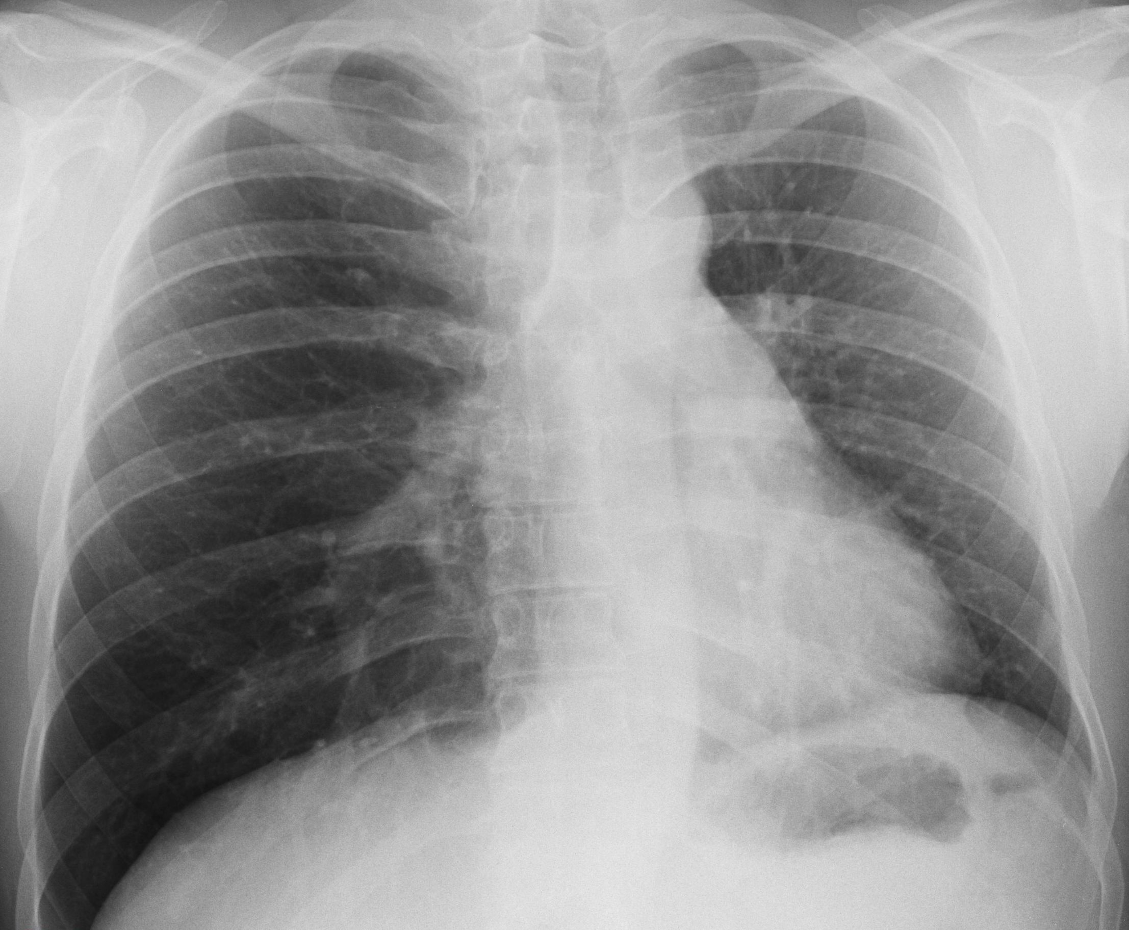
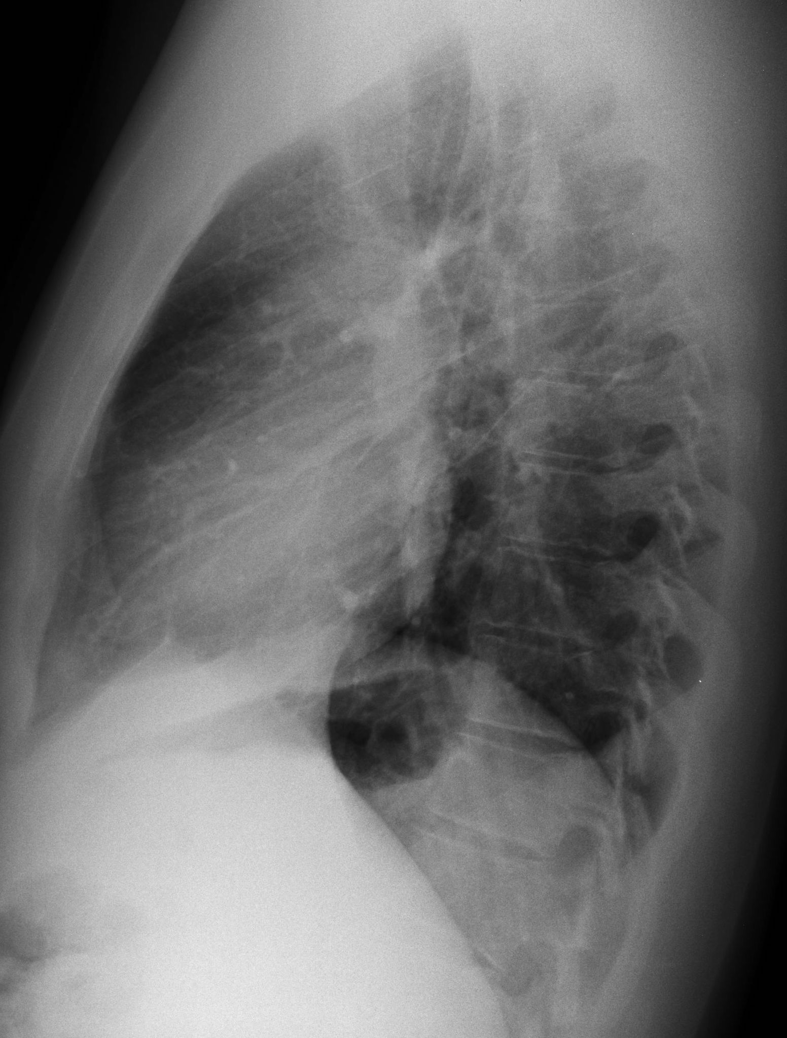
Today I am showing radiographs of a 30-year-old man with a persistent cough. What do you see?
Check the images below, leave your thoughts in the comments section and come back on Friday for the answer.
The main findings are seen in the PA radiograph, which shows displacement of the mediastinum towards the left. There is a discrete downward displacement of the left hilum (A, red arrow), as well as rotation of the cardiac silhouette causing loss of concavity of the left heart border (A, white arrow), the so-called flat waist sign, suggestive of LLL collapse. The findings are more evident when comparing with a previous film (B) taken two years earlier.

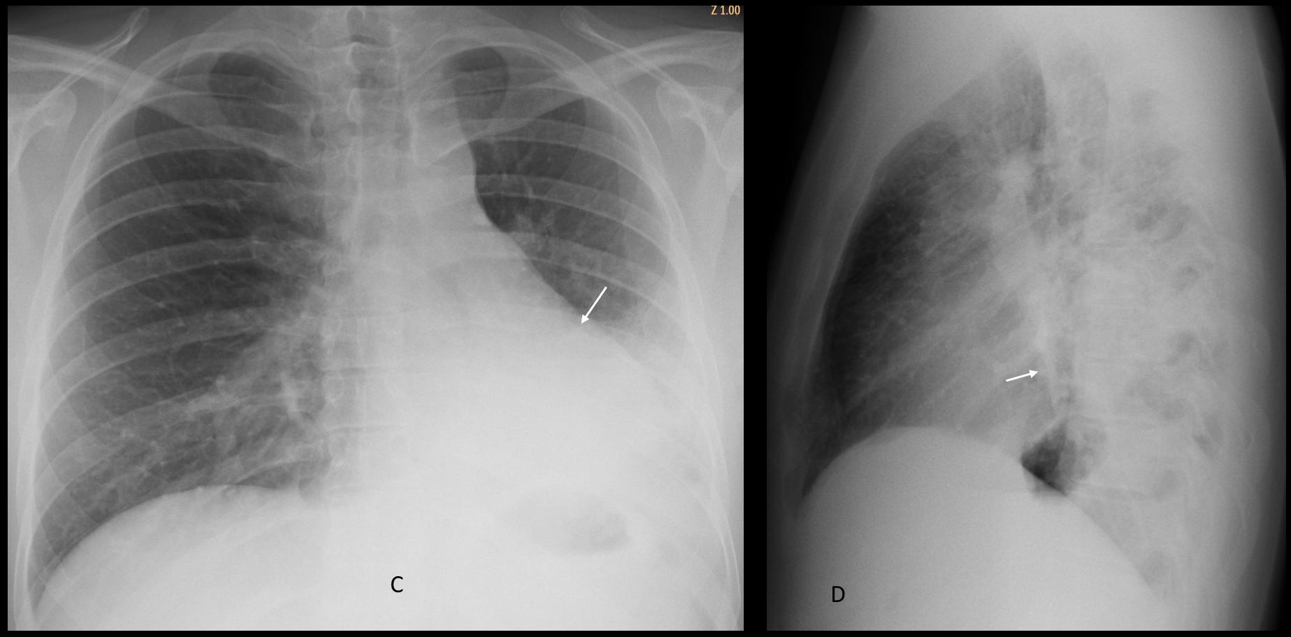
Findings were overlooked, and the study was read as normal. Five months later the patient returned with acute dyspnea. Chest radiographs show obvious signs of LLL collapse (C and D, arrows).
Unenhanced CT images show a rounded tumour in the left main bronchus (E-G, red arrows), confirmed to be a carcinoid tumour.
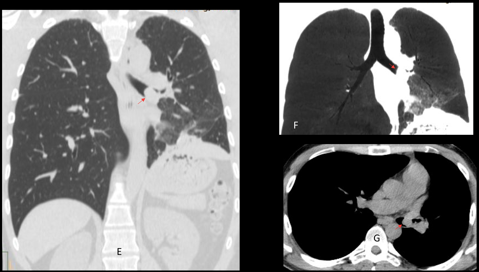
Final diagnosis: endobronchial carcinoid causing LLL collapse.
Congratulation to Beata who was the first to suggest the correct diagnosis.
Teaching point: remember to analyse the radiograph carefully in order to detect significant findings. In this case, several of you missed them and placed the abnormality on the right side, instead of on the left.