Dear Friends,
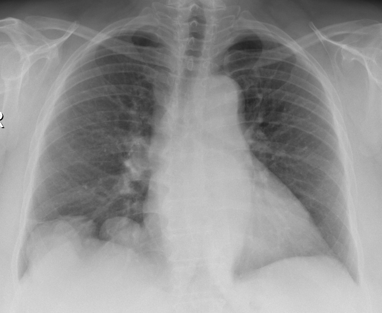
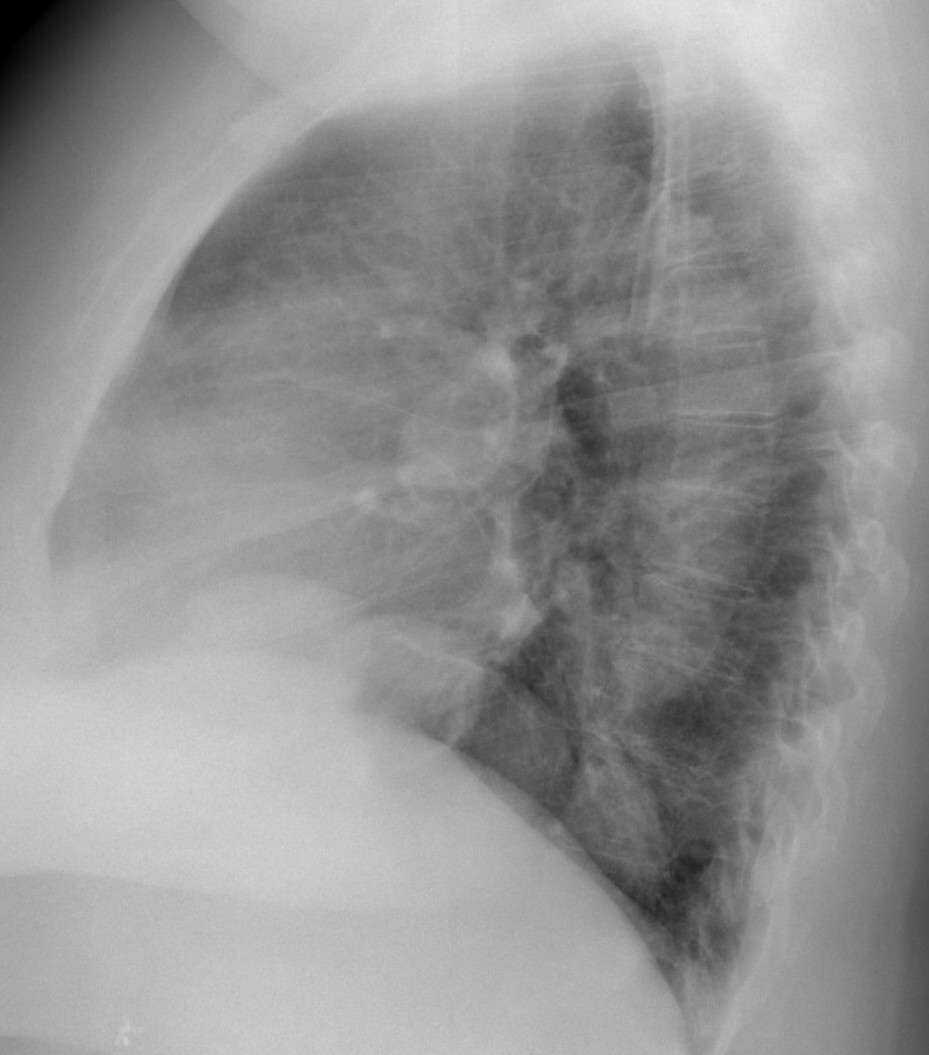
Today I am presenting a case lent to me by my good friend Dr. José Luis López Moreno. The images are preoperative radiographs of a 61-year-old woman with a pelvic mass. What do you see?
Check the images below, leave your thoughts in the comments section and come back on Friday for the answer.
Click here for the answer
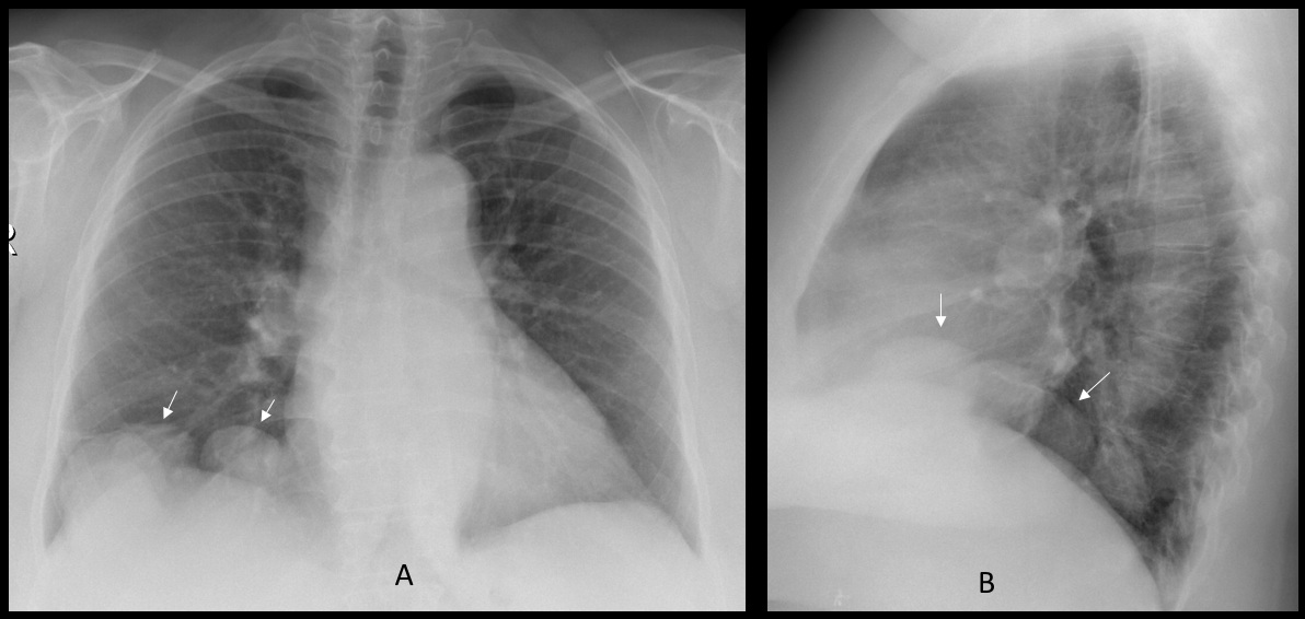
Findings: chest radiographs show several well-defined nodules at the right lung base (A-B, arrows). Considering that the patient has a pelvic mass, a likely diagnosis is metastases from an ovarian carcinoma.
Enhanced CTs of the right upper quadrant show that the nodules are in intimal contact with the liver and have similar density to the liver parenchyma (C-D, arrows). Coronal and sagittal enhanced CT demonstrate liver vessels going into one of the nodules (E-F, red arrows). A follow-up CT six months later (not shown) did not show any change.
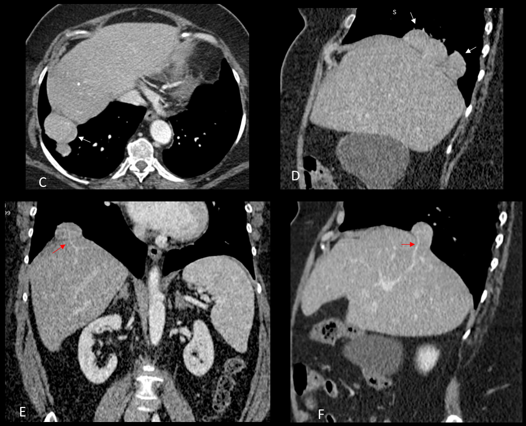
Final diagnosis: multiple liver hernias through the diaphragm.
This case is highly unusual (never seen it before). I did not expect you to make the diagnosis but hoped to be forgiven because of the beauty of the images.
Teaching point: remember that any apparent lung abnormality adjacent to the diaphragm may arise from below, as this case proves.


