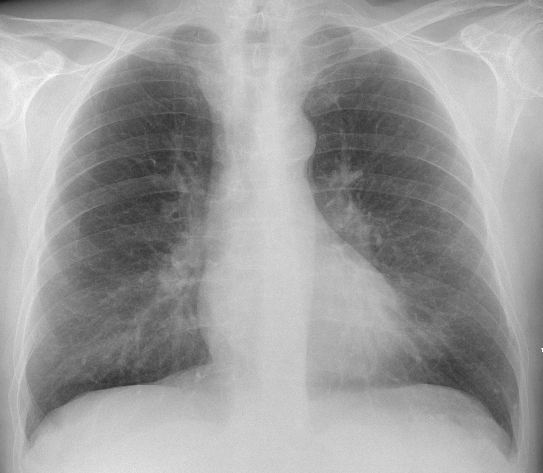
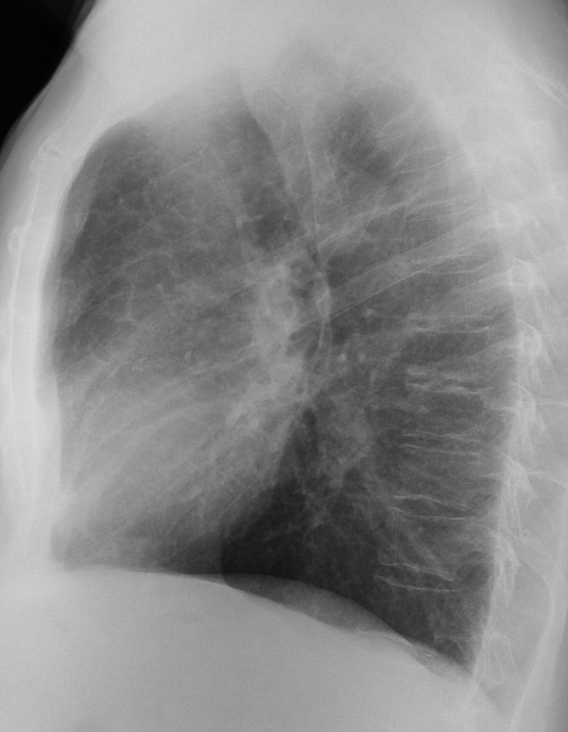
Today’s radiographs belong to a 70-year-old man with a chronic cough. What do you see?
Check the images below and leave your thoughts in the comments section. More images will be shown on Wednesday, followed by the answer on Friday.
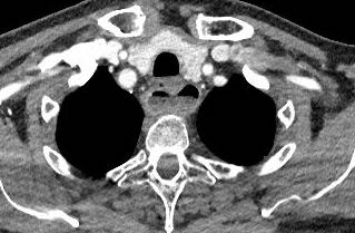
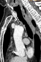
Dear Friends, now showing images of enhanced CT. Do they help?
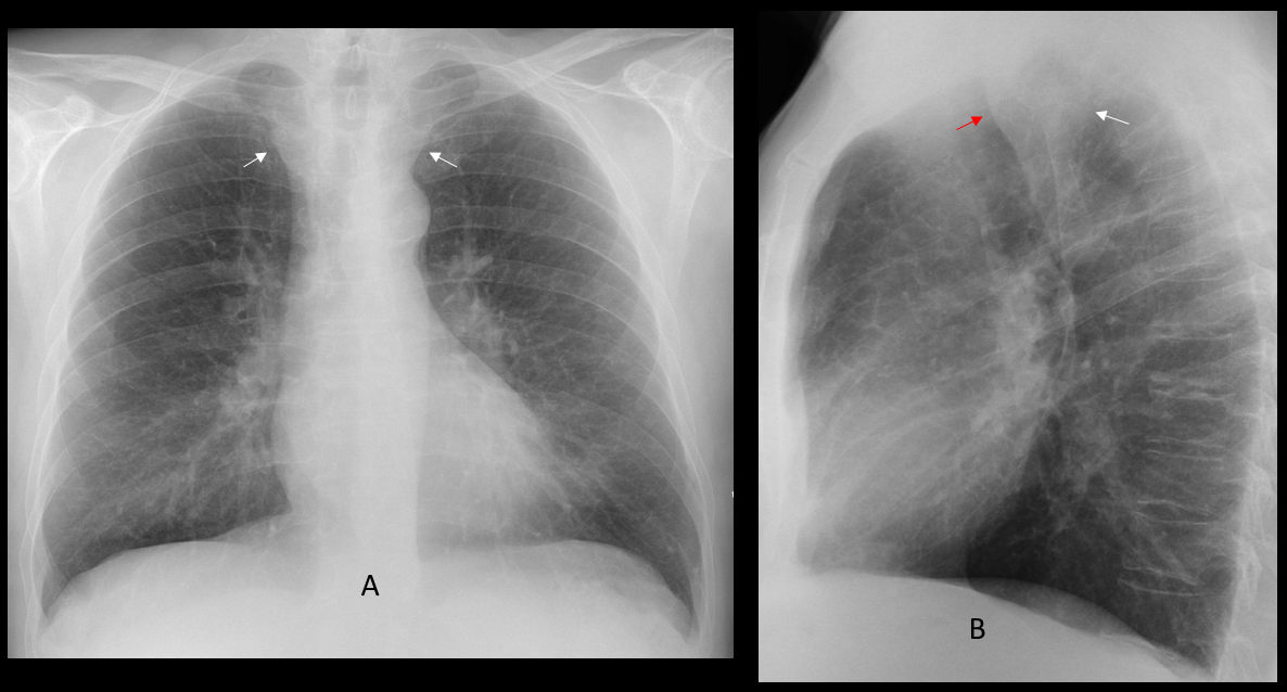
Findings: the PA radiograph shows a moderate widening of the upper mediastinum (A, arrows). The lateral view demonstrates a retro tracheal opacity in Raider’s triangle (B, arrow), bowing forward the posterior wall of the trachea (B, red arrow).
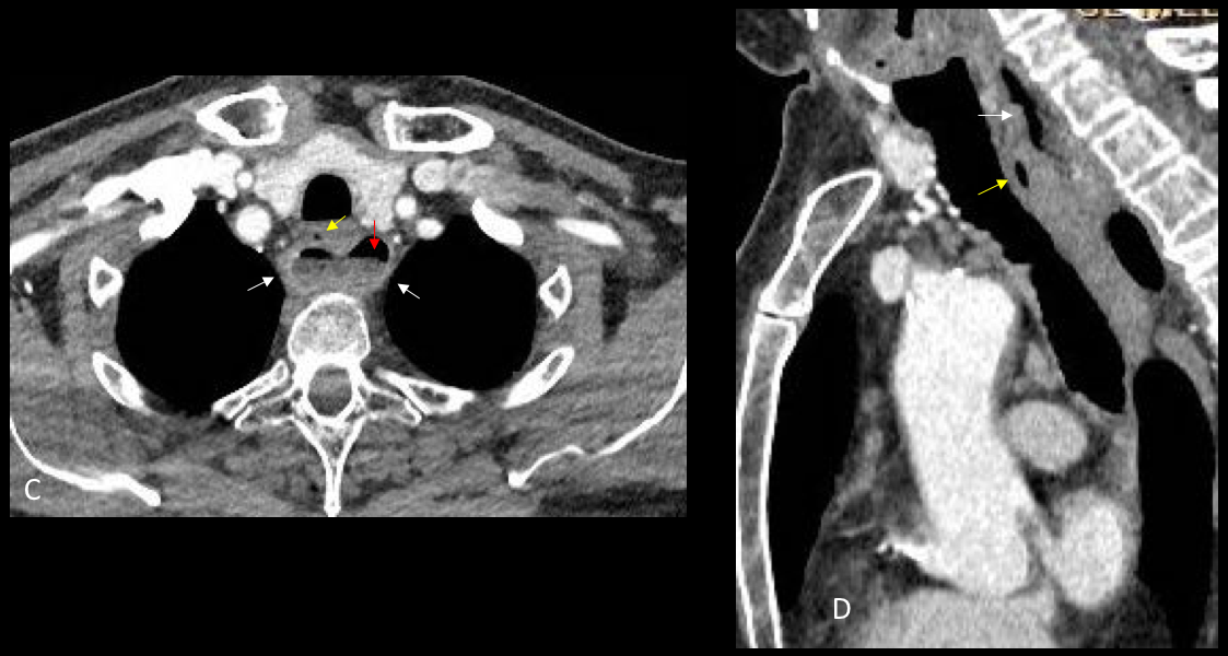
Enhanced axial and sagittal CT show a retro tracheal cavity (C-D, arrows) with an air-fluid level (C, red arrow). There is a bubble of air anteriorly (C-D, yellow arrows), which marks the oesophageal lumen.
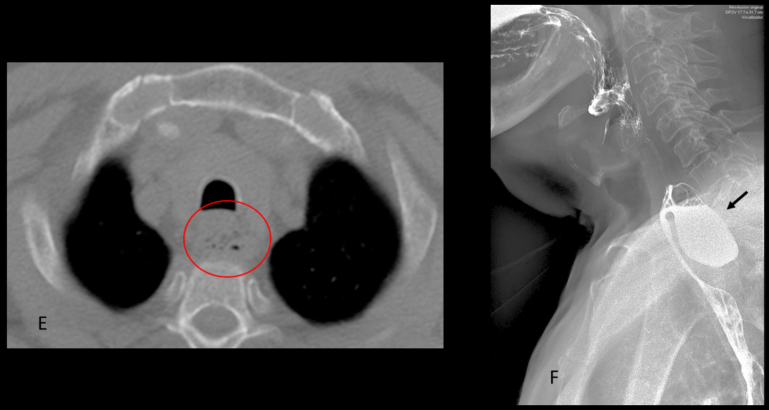
The findings suggested a Zenker diverticulum, which was confirmed with a barium oesophagogram (F, arrow). Review of a CT taken seven years earlier shows air bubbles in the same location (E, circle).
Final diagnosis: unsuspected Zenker diverticulum
Congratulations to tr, who made the diagnosis and to Pratiba, who was the first to detect the opacity in Raider´s triangle.
Teaching point: remember to look at Raider’s triangle in the lateral view. It may prove very rewarding, as happened in this case.


Distal traqueal stenosis.
There is a right paratracheal density on the frontal view. The lateral chest X-ray shows obliteration of the Raider’s triangle with indentation on the posterior trachea. Likely oesophageal dilation causing chronic cough due to recurrent aspiration.
Good evening sir. Ap view.There is retro cardiac silhouette seen on the right side. Increased size and density of the left hilum is also noted.
Lat view shows decreased vertebral body height of D3.
Bulla in the right lower zone, trachea and intermediate bronchus seem dilated and tortuos.
Greetings;
widening of the superior mediastinum on the PA view with obliteration of the retro-tracheal space on the lateral view with mild compression of the posterior wall of the trachea.
DDX of the retrotracheal space lesions:
1-vascular lesions/anomalies.
2-esophageal abnormalities.
3- mediastinal abnormalities (tumors, infections, and intra-thoracic goiter).
Good differential. Look at the CT tomorrow
Good moorning!
Distal trachea estenosis and right paratracheal line widening (probably adenopathies).
In the lateral view there is a protrusion on the trachea probably because of an esophageal lesion.
Perhaphs there is a double cardiac contour because of an increased of the left atrium (?).
Can see that you still have problems with the paratracheal line 🙂
Yes, It’s my torture
Bilateral prominent interstitial marking, mainly basaly with Kerley’s lines, both hilai are enlarged, double right heart border and splaying of carina, pictures of left atrial enlargement, mitral diseases.
obscured left heart border, suspected of lingular consolidation.
sarcoidosis
? Aberrant right subclavian artery aneurysm causing right paratracheal density and posterior tracheal bulging (Kommerell’s diverticulum)
Greetings,
an air-fluid level can be seen posterior to the trachea, a small air bubble anterior to the this lesion probably represents the normal esophageal lumen, no abnormalities noted in the spine or para-spinal soft tissue.
impression: zenker diverticulum.
Achalasia cardia
Hi,
Thanks for sharing this information. There are some conferences happening in which medical specialty would be “Thoracic Radiology” and here is one of those conferences the conference details are given below.
Penn Radiology CME is Organizing Pearls, Pitfalls & Protocols: Spring Imaging Update 2020 held from Mar 23 – 27, 2020 at Lake Buena Vista, Florida, USA.
Source: https://www.emedevents.com/c/medical-conferences-2020/pearls-pitfalls-and-protocols-spring-imaging-update/
Hi,
Thanks for sharing this information. There are some conferences happening in which medical specialty would be Thoracic Radiology and here is one of those conferences the conference details are given below.
Diagnostic Imaging CME: Penn Radiology CME Organizing 42nd Annual Diagnostic Imaging: Back to the Vineyard held from Jul 06 – 10, 2020 at Edgartown, Massachusetts, USA.
For more information please follow the below link:
https://www.emedevents.com/c/medical-conferences-2020/42nd-annual-diagnostic-imaging-back-to-the-vineyard