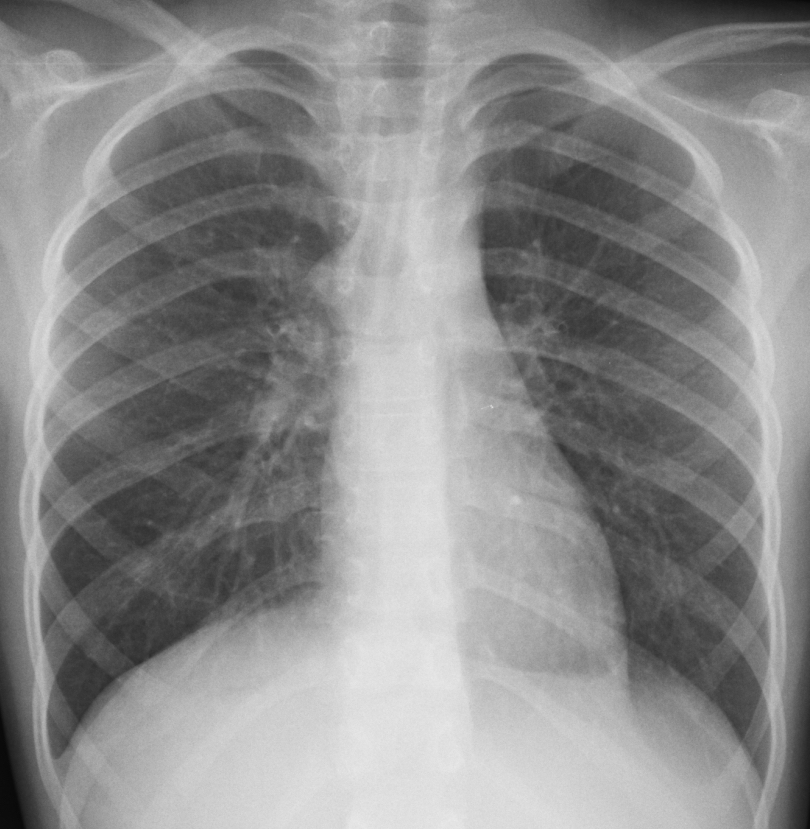
Presenting today chest radiographs of a 9-year-old girl with a persistent cough for the last two weeks, vomiting and fever. What do you see?
Check the images below and leave your thoughts in the comments section. More images will be shown on Wednesday, followed by the answer on Friday.
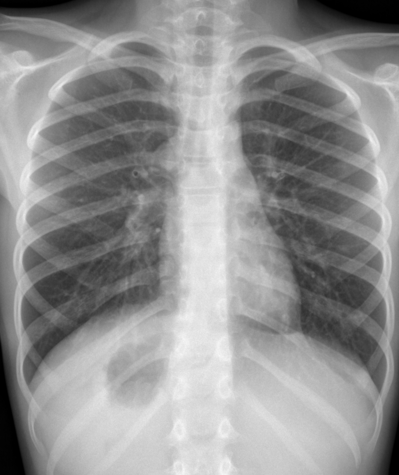
Dear friends, showing a chest radiograph taken seven days later. What do you see?
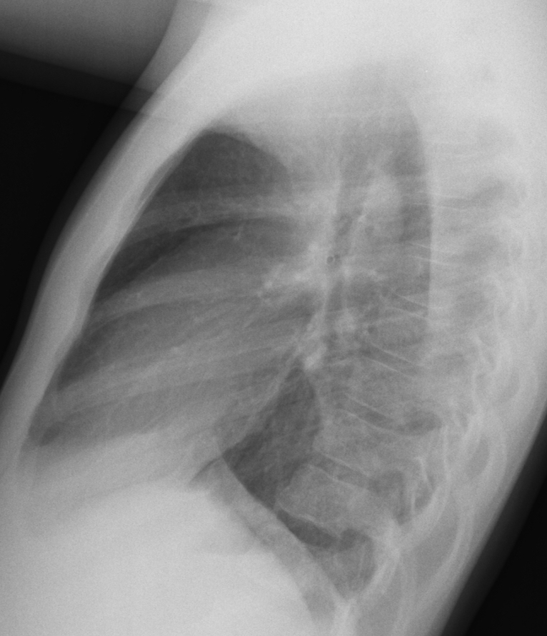
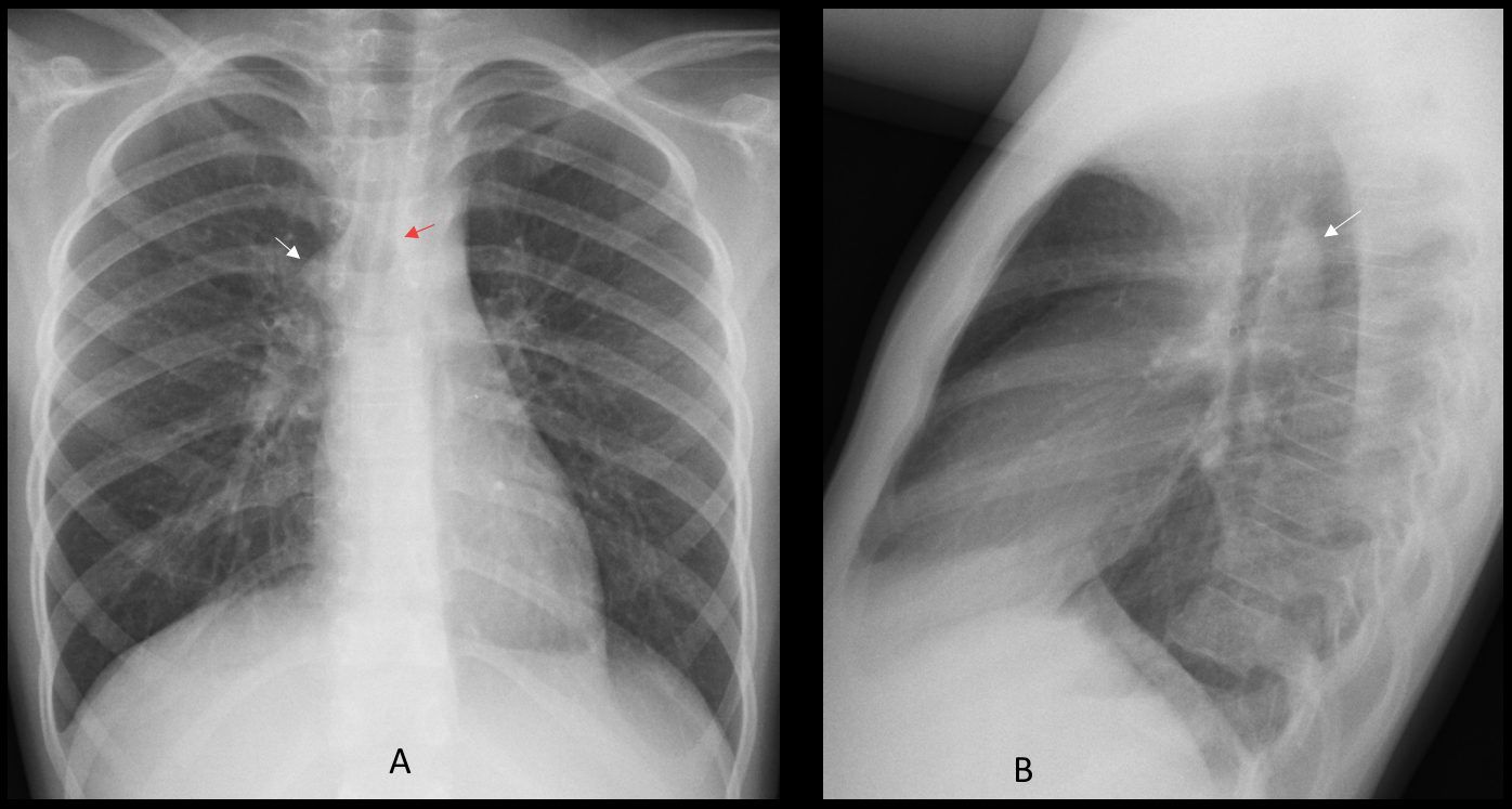
Findings: PA radiograph shows a rounded right mediastinal prominence at the level of the tracheal bifurcation (A, arrow). It could be either a lymph node or an enlarged azygos vein. The retrotracheal location in the lateral view (B, arrow) points to an azygos vein.
The aortic arch is located in the left side, as evidenced by the discrete imprint in the left tracheal wall (A, red arrow).
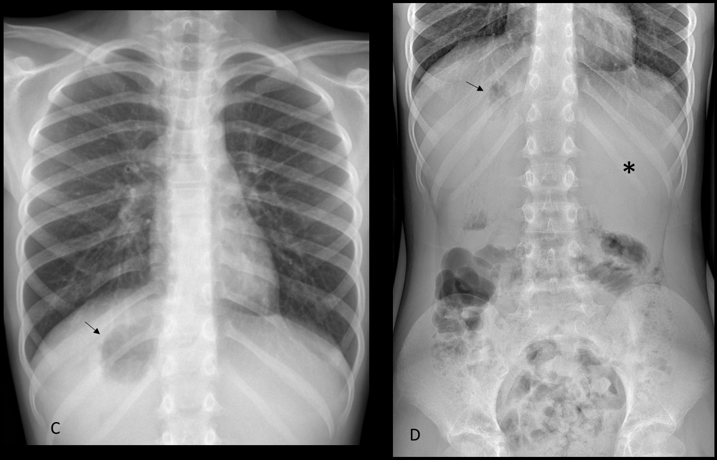
PA radiograph taken one week later demonstrates air in the gastric fornix, which is located under the right hemidiaphragm (C, arrow). It is confirmed by a supine film of the abdomen (D, arrow), which also shows the liver in the left upper quadrant (D, asterisk).
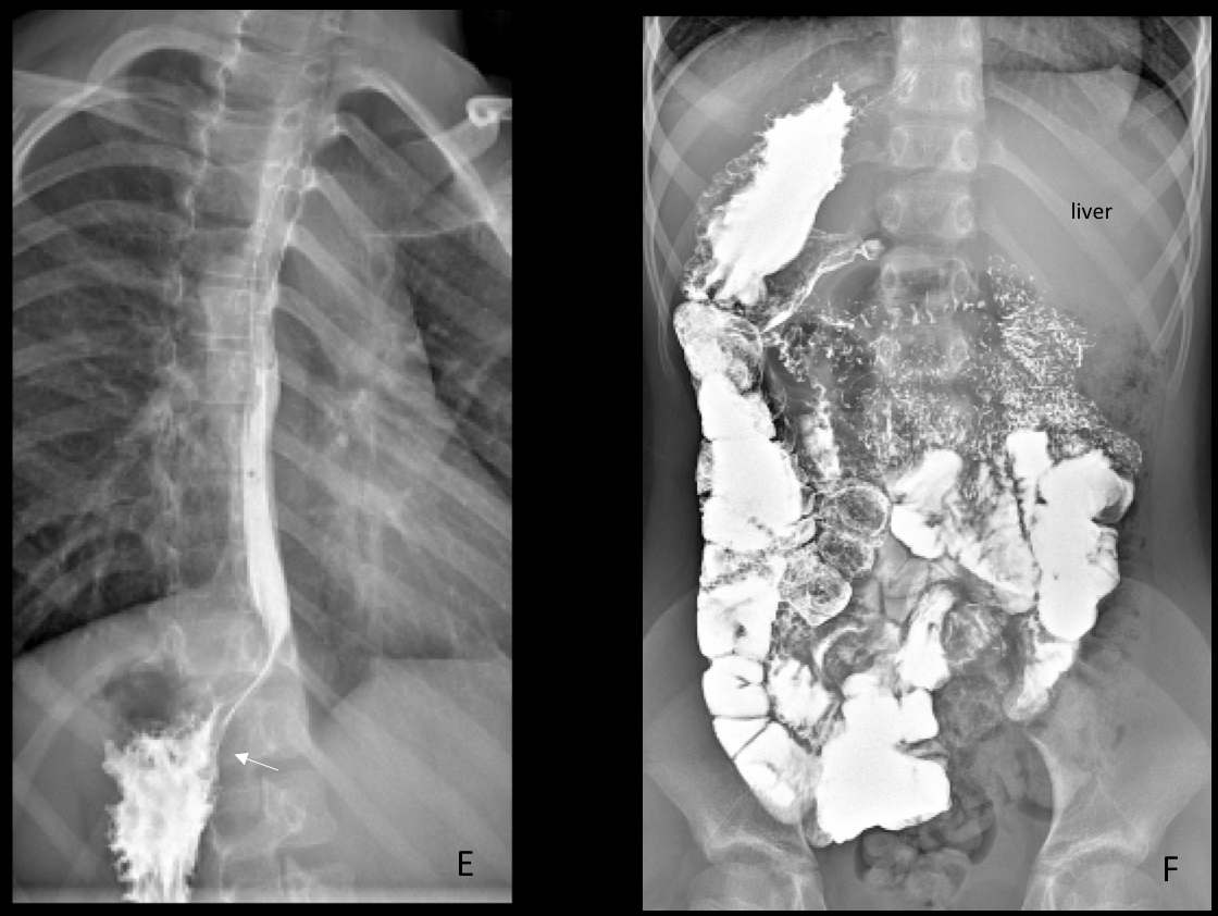
Barium study confirms the right-sided stomach (E, arrow) and malrotation of the bowel, mostly located in the right side of the abdomen (F).
The patient was diagnosed with whooping cough with positive serology to Bordetella Pertussis. The isolated abdominal malrotation was an incidental finding
Final diagnosis: Levocardia with abdominal heterotaxia and azygos continuation of IVC
Congratulations to MK and S who were the first to make the definitive diagnosis.
Teaching point: Sometimes, the unusual is more usual than expected. This is our third patient with levocardia and abdominal heterotaxia seen in the last four years (see Caceres´ Corner Case 194 and Dr. Pepe´s Summer Case 1). It may not be as rare as the textbooks state!