Dear friends,
Showing post-operative radiographs of a 34-year-old male. Muppet saw the films without clinical information and was completely lost.
Can you interpret the findings and shame Muppet into early retirement? Check out the images below, post your thoughts and possible diagnoses in the comments section, and come back next Tuesday for the answer!
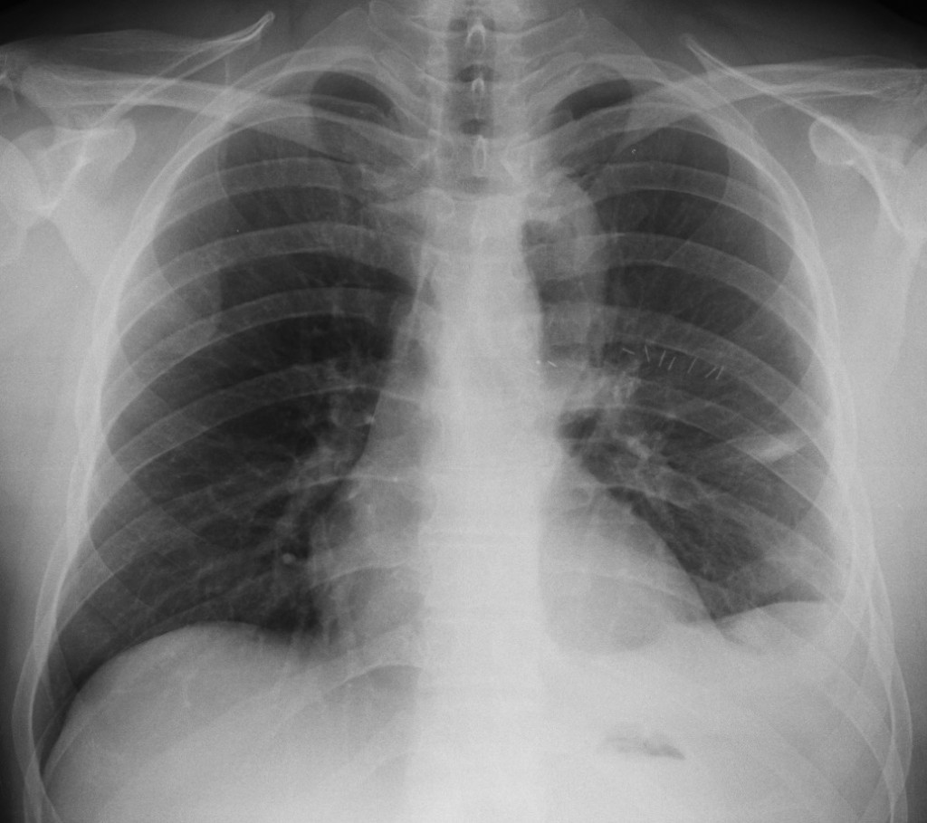
34-year-old male, PA chest
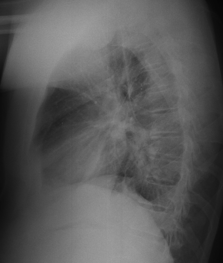
34-year-old male, lateral chest
Click here for the answer to case #72.
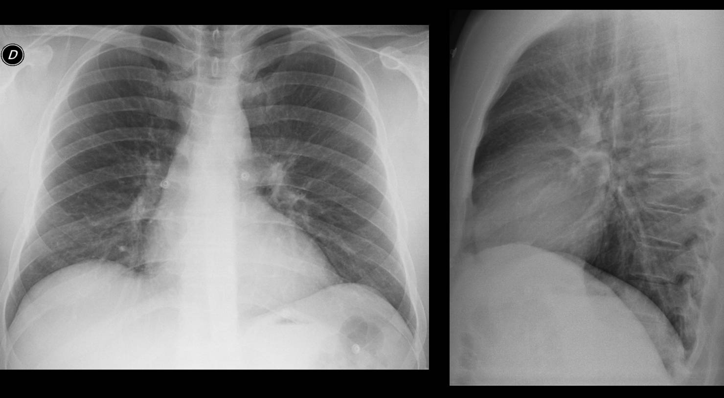
Findings: chest radiographs show post-operative changes in the left hemithorax, with surgical clips in the mid-field and residual pleural effusion. There is a para-mediastinal tubular structure, better seen in the immediate post-operative film (A, B, arrows). As the majority of you mentioned, a tubular structure in the lungs suggests a vascular lesion. Muppet investigated the clinical history and it corresponds to a graft between the subclavian artery and descending aorta in a patient with aortic coarctation. The surgeon found a hypertrophic Adamkiewicz artery with marked collateral circulation and was afraid to resect the coarctation and cause damage to the spinal cord (thus the graft).
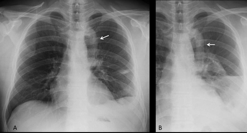
Fig. 1
Muppet checked the pre-op films (Fig. 2) and there were no imaging findings suggestive of coarctation: no rib notching, non-prominent ascending aorta and unremarkable aortic knob. This normal appearance may be related to collateral circulation going through Adamkiewicz artery.
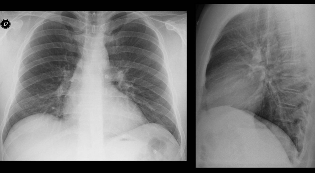
Fig. 2
Final diagnosis: aortic coarctation corrected with bypass graft.
Teaching point: tubular images in the lung should suggest three possibilities: vascular, bronchial impaction and herniated bowel loops.