Dear Friends,
Presenting the last case before Summer vacation. Muppet needs quality time to replenish his energy (July and August) and will return in September with more interesting (and teaching) cases.
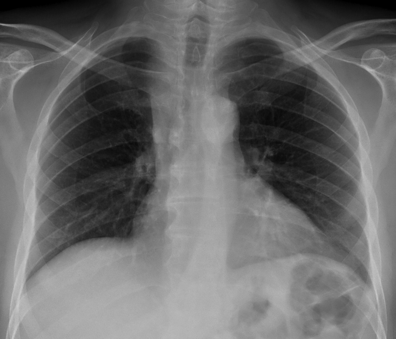
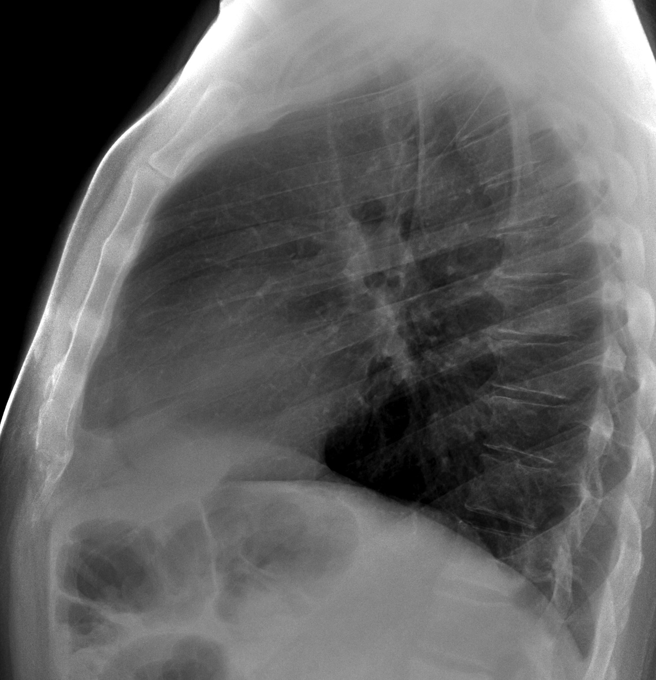
These radiographs belong to a 52-year-old male with a cough. The answer has now been added, but you can still leave your thoughts and diagnosis in the comments section, below, before you check it.
Diagnosis:
1. Mediastinal fat
2. Dilated SVC
3. Lymphoma
4. None of the above
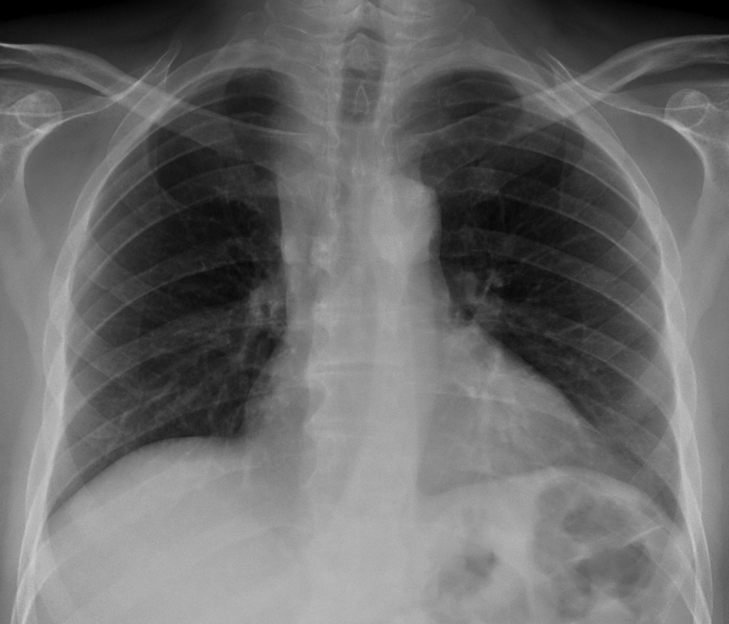
52-year-old male, PA chest
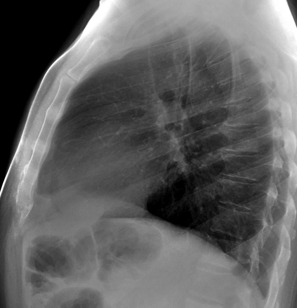
52-year-old male, lateral chest
Click here for the answer to case #72.
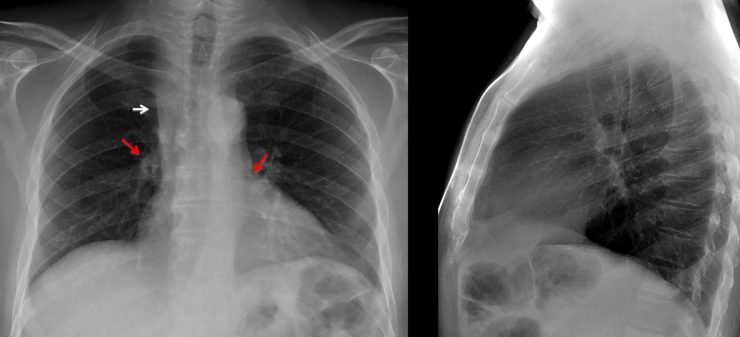
Findings: PA chest shows widening of the right upper mediastinum (arrows). The first diagnosis that comes to mind is enlarged lymph nodes. Other possible causes are an enlarged vena cava or mediastinal fat. The clue to the diagnosis lies in the right hilum, which is higher than the left (
red arrows).
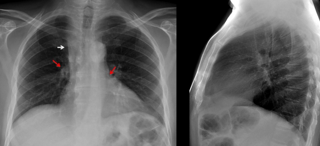
At no time should this occur, which raises the possibility of loss of volume of RUL pulling up the right hilum. Coronal CT shows marked collapse of RUL which is visible as a thin stripe apposed to the mediastinum (A, arrows). Coronal reconstruction shows the obstructed bronchus (B, yellow arrow) leading into the collapsed RUL (arrows).
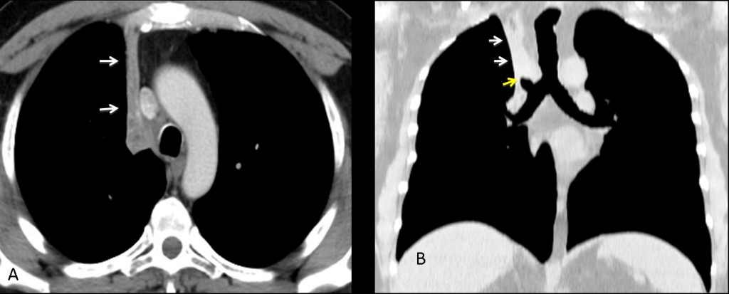
Final diagnosis: marked RUL collapse, secondary to bronchial carcinoma.
Congratulations to Alice, who suggested the correct diagnosis.
Teaching point: do look at the hila. They may provide important information.
Muppet wishes you all a pleasant vacation!