Dear Friends,
The Muppet is very happy with your responses to the previous case. As a prize, he wants to show you the following case that we saw together this July before he went on vacation with Miss Piggy (and left me working). Radiographs belong to a 46-year-old woman, with pain in the chest.
Leave your thoughts and diagnosis in the comments section below, and come back on Friday for the answer.
Diagnosis:
1. Empyema necessitatis
2. Chest wall tumour
3. Metastatic disease
4. None of the above

46-year-old woman, PA chest
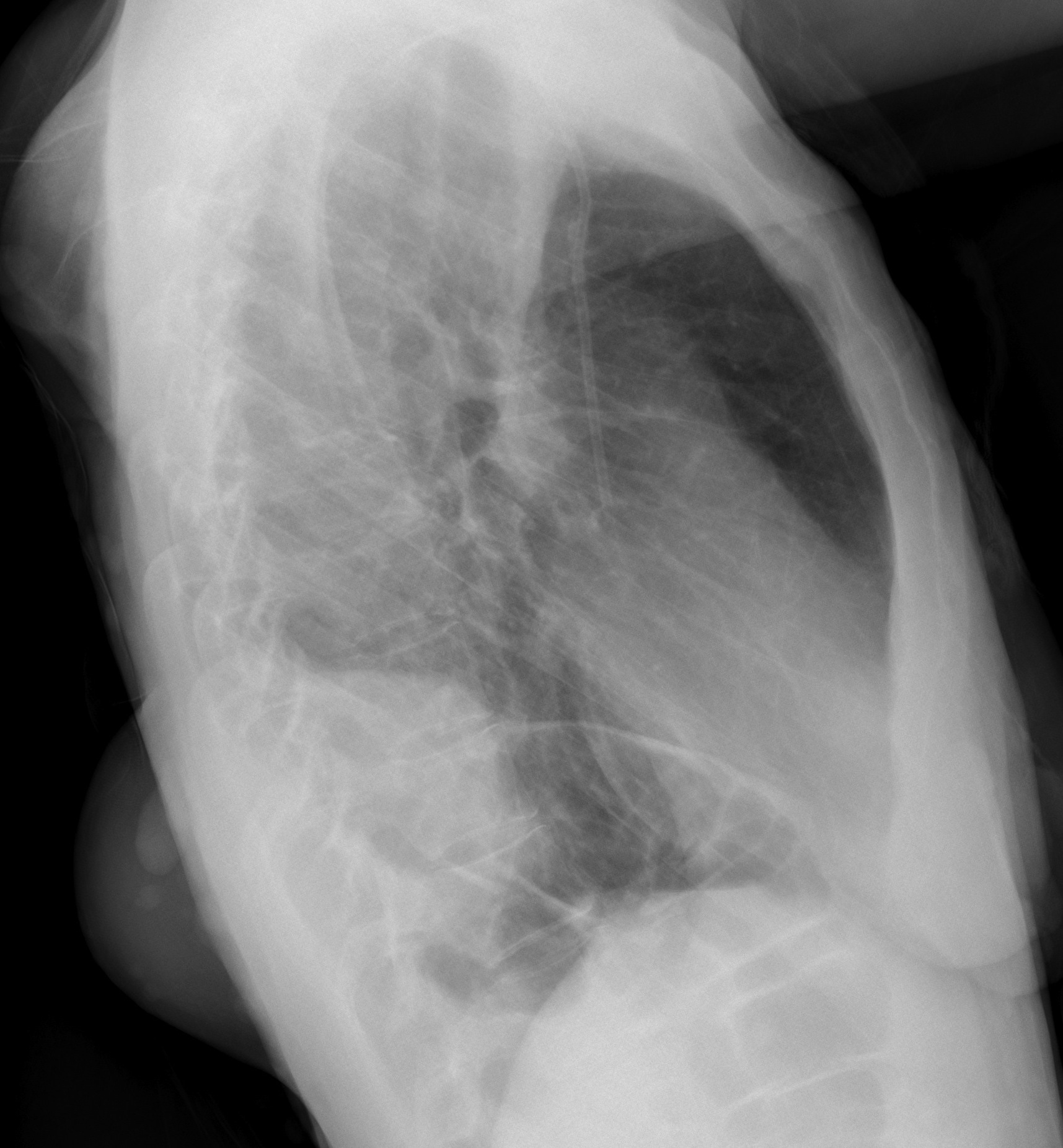
46-year-old woman, lateral chest
Click here for the answer to case #75.
Findings: lateral chest shows an inferior-posterior mass invading the soft tissues of the chest wall (arrows). There is a false mass above, which corresponds to the scapula (
yellow arrow). The PA view barely demonstrates the lower mass, hidden behind the diaphragm (arrow).
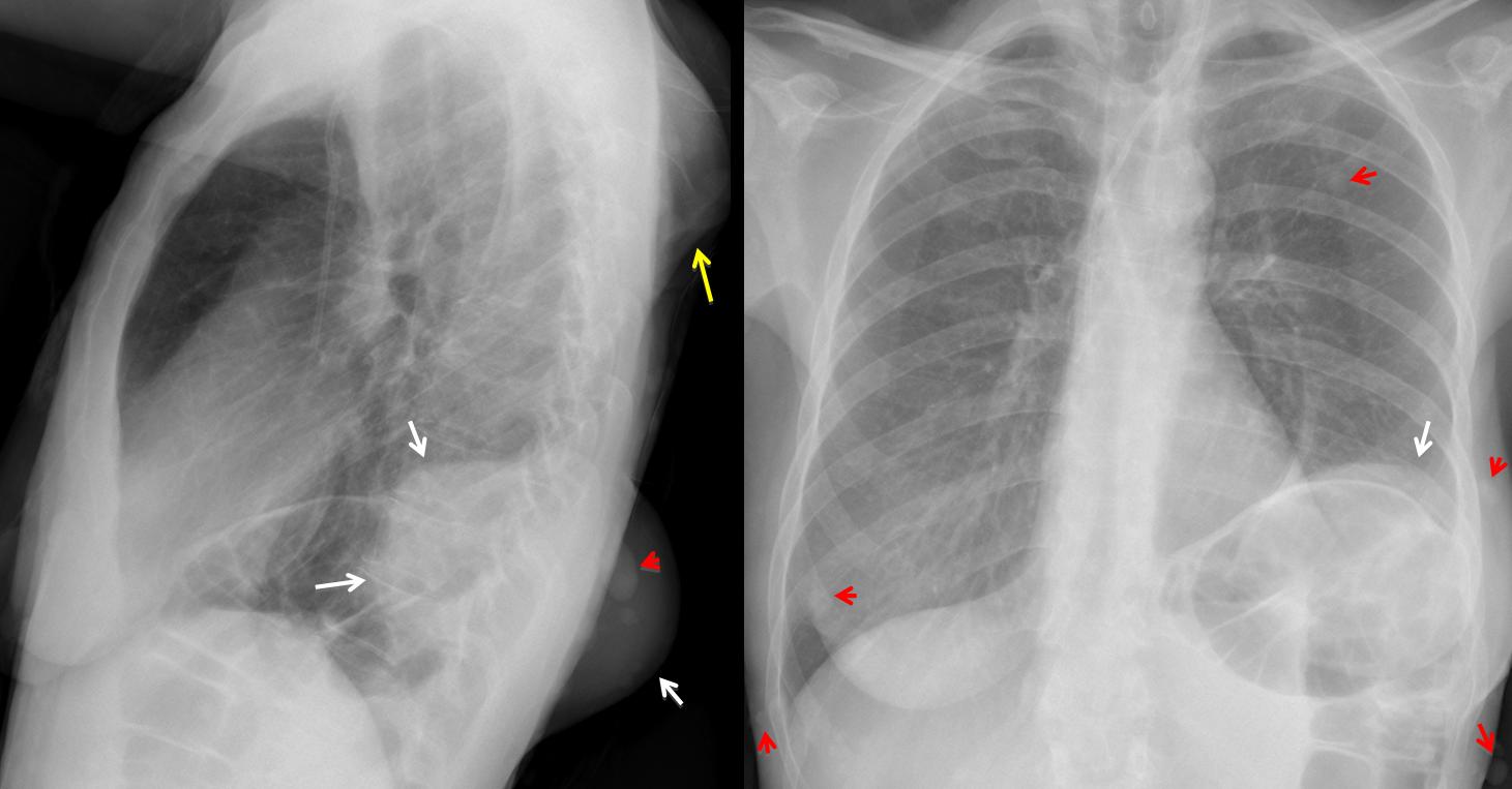
Fig. 1
The clue to the diagnosis lies in the numerous soft tissue nodules (red arrows), some of them projected over the lungs, simulating lung metastases. Findings are confirmed with CT (Fig. 2, arrows). This combination of skin nodules and a large chest wall mass is highly suggestive of neurofibromatosis with associated tumour.
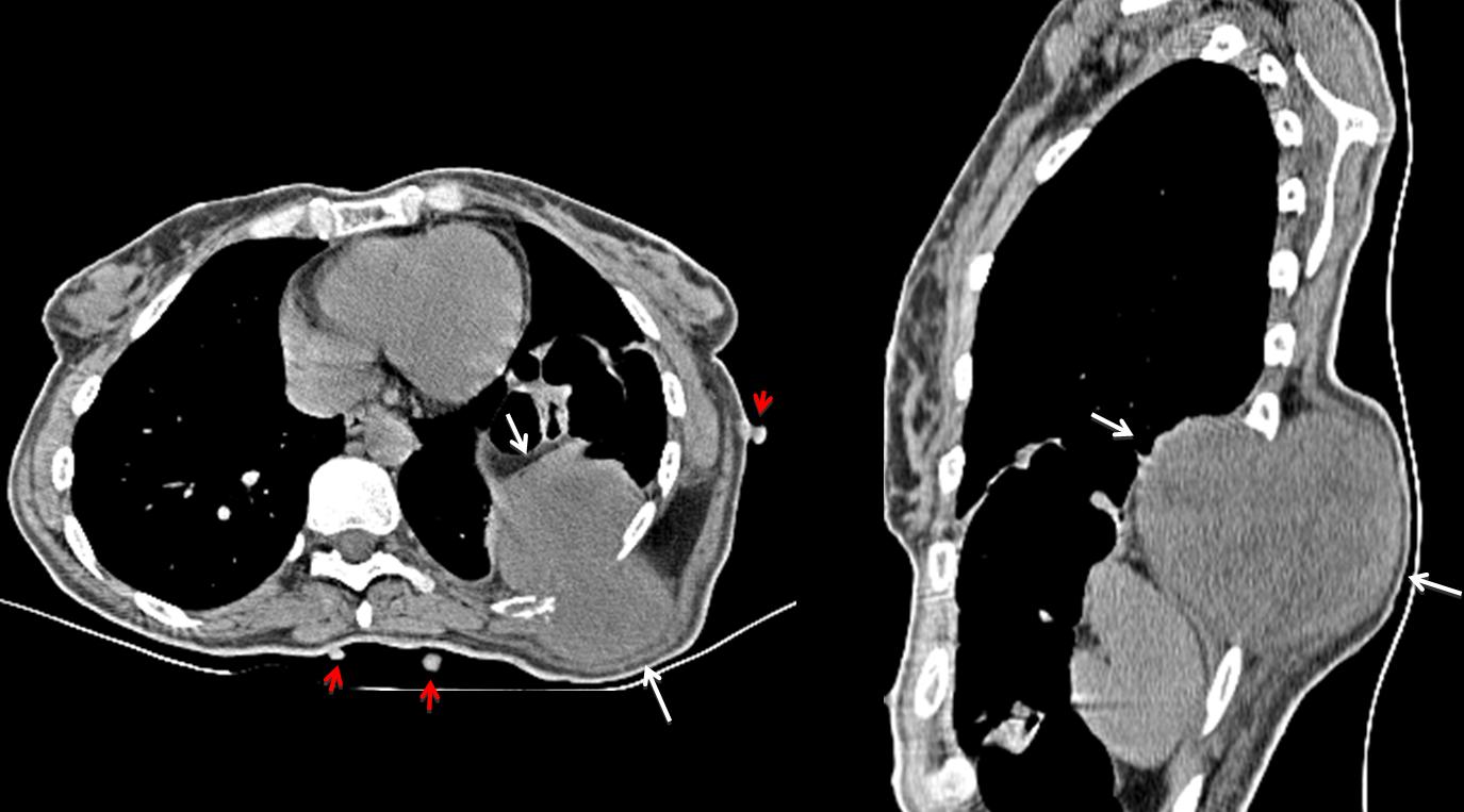
Fig. 2
Final diagnosis: Neurofibromatosis with neurofibrosarcoma of the chest wall.
Congratulations to Gus, who was the first to answer and to suggest the correct diagnosis.
Teaching point: always look beyond the obvious. Subtle findings may point towards the right diagnosis.


