This week Muppet and I are going on a dangerous trip abroad. Forgive me if I answer your comments a little bit later than usual; the locals may be hostile and refuse to share their WiFi.
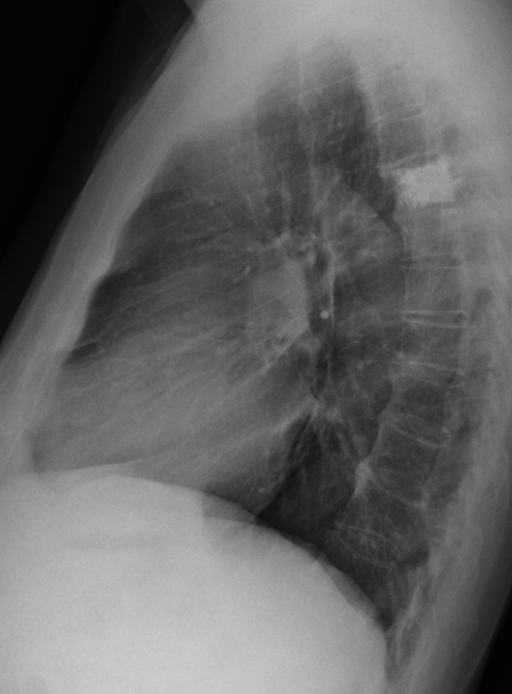
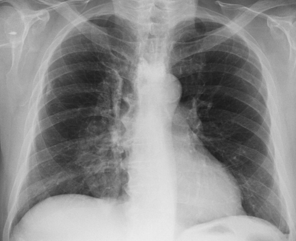
Radiographs belong to a 73 y.o. male with fever and symptoms of acute prostatitis.
The obvious finding is the increased density of the fifth dorsal vertebra (Fig 1, arrows). The vertebral body has ragged borders with metallic opacities within and it’s smaller than the ones above and below (insert). The initial impression is osteoblastic metastases, supported by a clinical history of prostatic disease. However, the metallic opacities and the shortening of the vertebral body suggest a previous surgical procedure. Review of the clinical history revealed that surgery for a vertebral hemangioma had been performed several years ago (probably vertebroplasty).
There is also a right paramediastinal calcified ovoid opacity in the PA view, barely visible in the lateral view as a posterior dense line (Fig 1, red arrows).
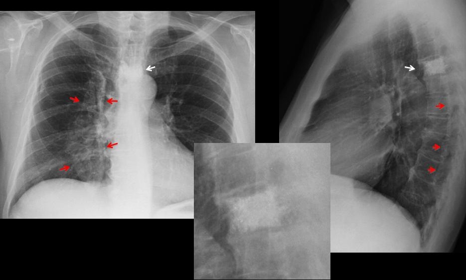
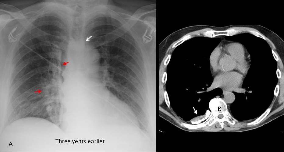
It has the appearance of pleural calcification, confirmed by upper slices of an abdominal CT performed at the time of admission (Fig 2B, arrow). Patient had a history of pneumothorax in his youth.
A previous AP radiograph three years earlier confirms that the pleural opacity and the dense vertebra were present and unchanged (Fig 2A, arrows).
Final diagnosis: Iatrogenic dense vertebra (probable vertebroplasty). Unrelated pleural calcification.
Congratulations to Katherina, who was the first to make the correct diagnosis.
Teaching point: always remember that iatrogenia may cause any type of finding, especially when metallic opacities are visible. And remember that all visible findings do not have to be attributed to a single disease.