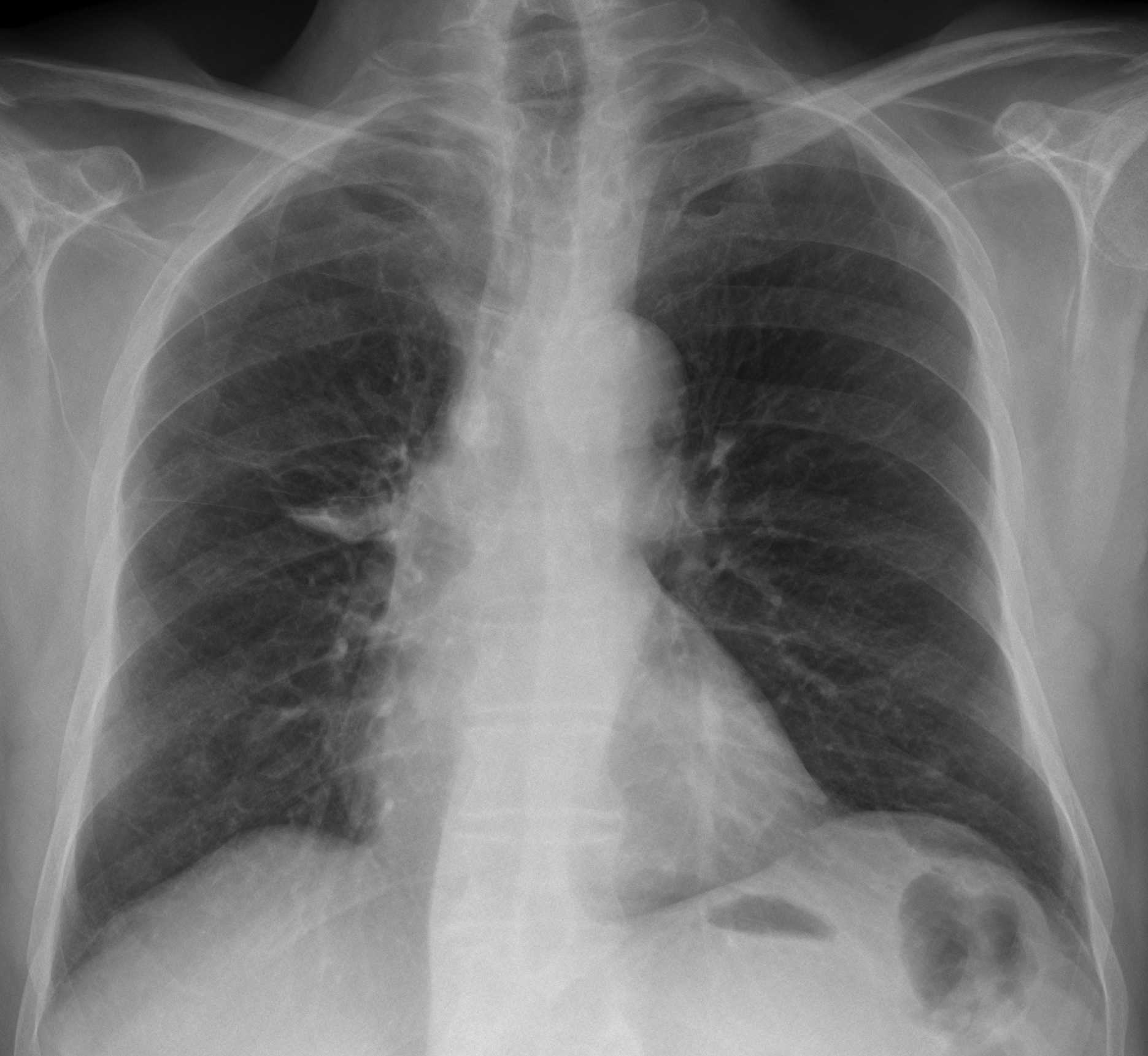
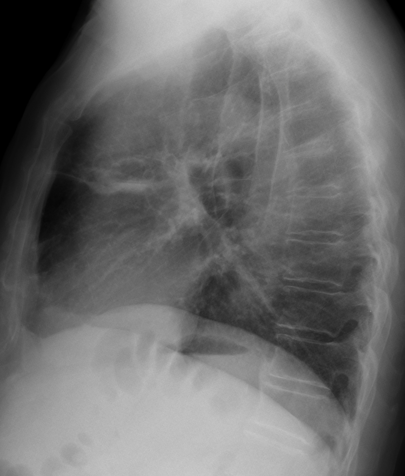
This week I am presenting radiographs of a 78-year-old male with haemoptysis. Have a look at the images below and leave us your thoughts and diagnosis in the comments section. The answer will be added on Friday.
1. Hydropneumothorax in minor fissure
2. Tuberculosis
3. Carcinoma
4. None of the above
Findings: PA radiograph show a cystic RUL lesion with thin walls and an air-fluid level. On the lateral view there are several cystic cavities, at least two of them with air-fluid levels (Fig. 1, arrows).
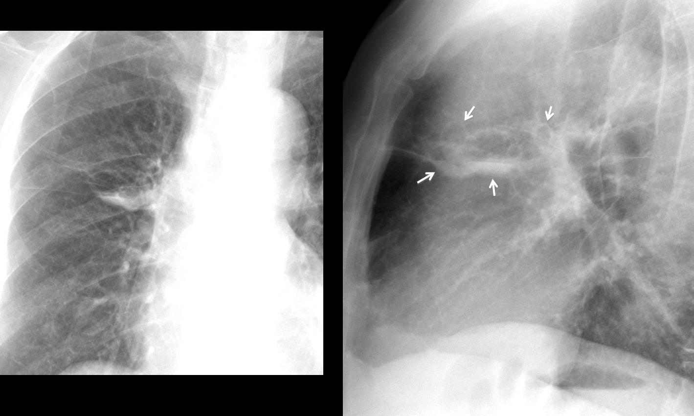
Fig. 1
The thin walls and the multiplicity rule out carcinoma and it is obvious that some lesions are outside the minor fissure, which excludes option one. The radiographic appearance suggests bronchiectasis, confirmed with axial CT (Fig. 2a, arrows) performed two years earlier. At that time, bronchiectasis were partially fluid-filled (Fig. 2b arrow), simulating a pulmonary nodule in the chest radiograph (Fig. 3, arrows).
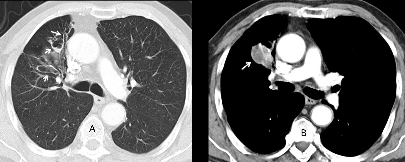
Fig. 2
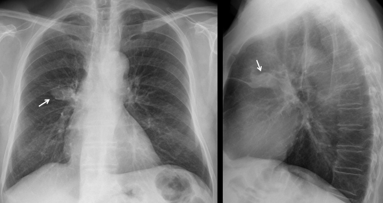
Fig 3. Two years earlier
Congratulations to Gus, who was the first to suggest the diagnosis, with Marcy as a close second.
Final diagnosis: cystic bronchiectasis, probably secondary to previous TB (not confirmed).
Teaching point: pay attention to details. In this case, the presence of several thin-walled cavities in the lateral view helped to rule out some of the proposed diagnoses and suggested the right one.