
Dear Friends,
Muppet saw this case while looking at daily chest radiographs and it caught his attention. We looked at the patient’s history and found that she was a 64-year-old female with back pain, who had a well-differentiated liposarcoma removed from her right thigh seven years earlier.
Do you see anything?
What do you think it is?
Check the images below and leave your thoughts in the comments section. The answer will be posted on Friday.

64-year-old female, PA chest

64-year-old female, lateral chest
Click here for the answer to case #78
Findings: the PA radiograph shows a double contour of the aortic knob (arrows). It does not obliterate the aortic contour, indicating a mediastinal mass in front of or behind the aorta. The lateral view shows increased opacity over the middle vertebra (circle), suggesting a posterior location.
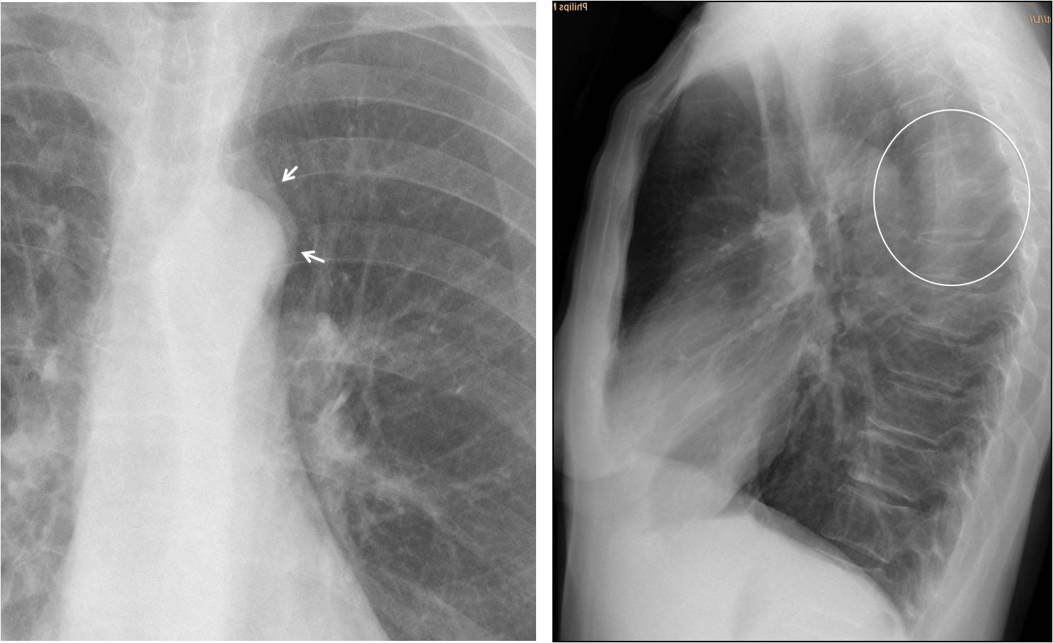
Fig. 1
Coronal and axial CT demonstrate a posterior mediastinal soft tissue mass (arrows). Without the clinical history I would have suspected a neurogenic tumour. However, MRI shows involvement of the adjacent vertebra, making metastases more likely.

Fig. 2
Final diagnosis: vertebral metastases from liposarcoma, surgically proven.
Congratulations to Martita, who made a brilliant diagnosis. Muppet elated!
Teaching point: it is my impression that the majority of us look mainly at the lungs and forget about the remaining structures of the chest (not Muppet, though). So, remember to look carefully at the whole chest. This case was discovered because the plain radiograph looked abnormal.


