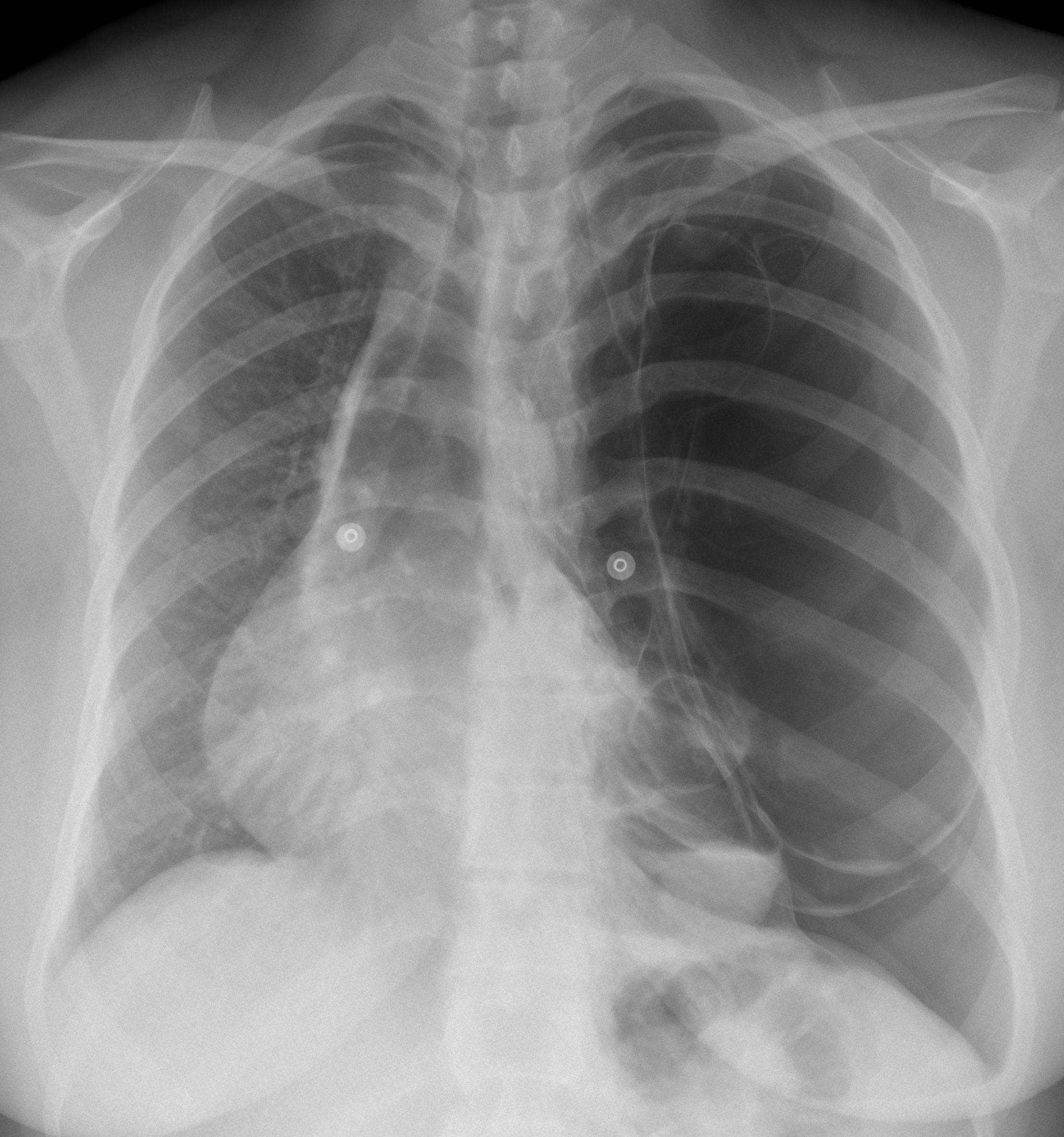
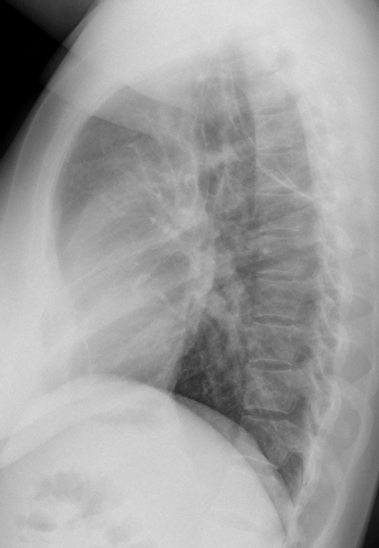
This week I am showing you a case provided by my good friend Dr. Jordi Andreu. The radiographs below belong to a 39-year-old woman with increased shortness of breath for the last three months. Leave your thoughts and diagnosis in the comments section and come back for the answer on Friday.
1. Bullous emphysema
2. Tension pneumothorax
3. Adenomatoid malformation
4. None of the above
Findings: PA chest shows an expanded hyperlucent left lung with displacement of the mediastinum towards the opposite side. The left hemithorax is occupied by several thin-walled cavities (arrows), one of them with an air-fluid level (
red arrow). The lateral view does not provide any additional information.
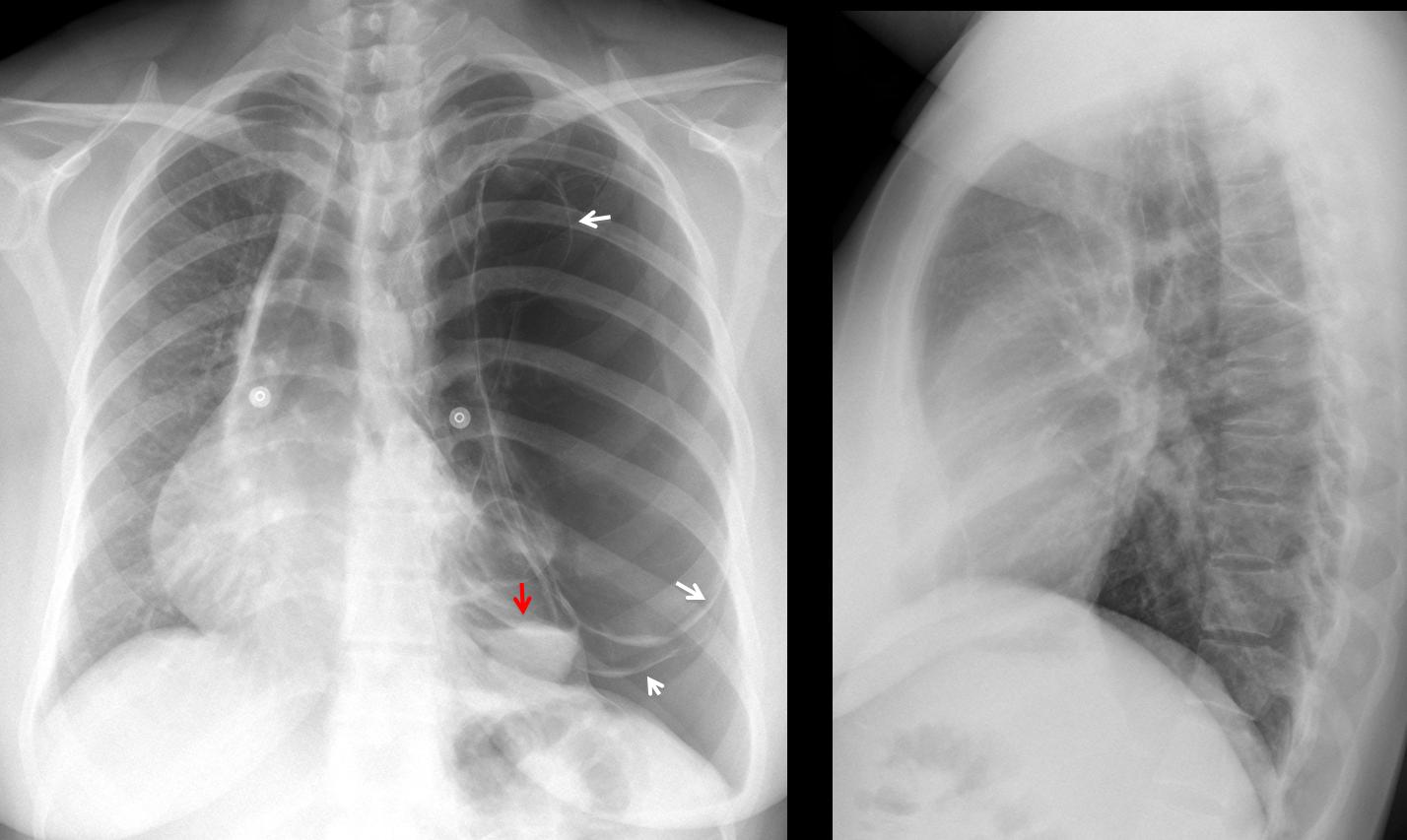
Fig. 1
Axial and coronal CT show several thin-walled large bullae; one of them with air-fluid level (Fig 2, red arrow). There is a small pneumothorax with some fluid as well. The left lung is collapsed (ryellow arrows). The right lung looks normal.
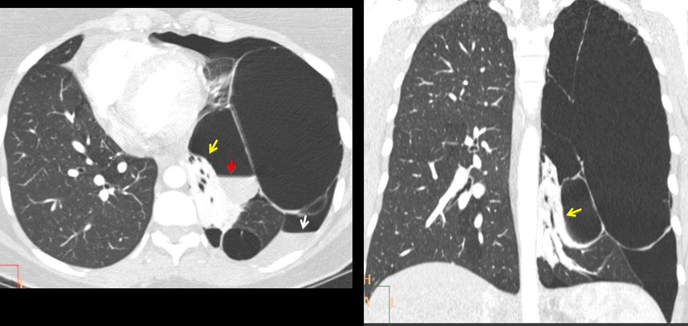
Fig. 2
This is a difficult case and the main differential diagnosis is between giant bullous emphysema and congenital cystic malformation. I consider tension pneumothorax very unlikely because the walls of the bullae are rounded and not flattened. The patient was operated on and the final diagnosis was unilateral giant bullous emphysema (vanishing lung syndrome). Post-op radiograph shows a near-normal appearance of the chest (Fig. 3).
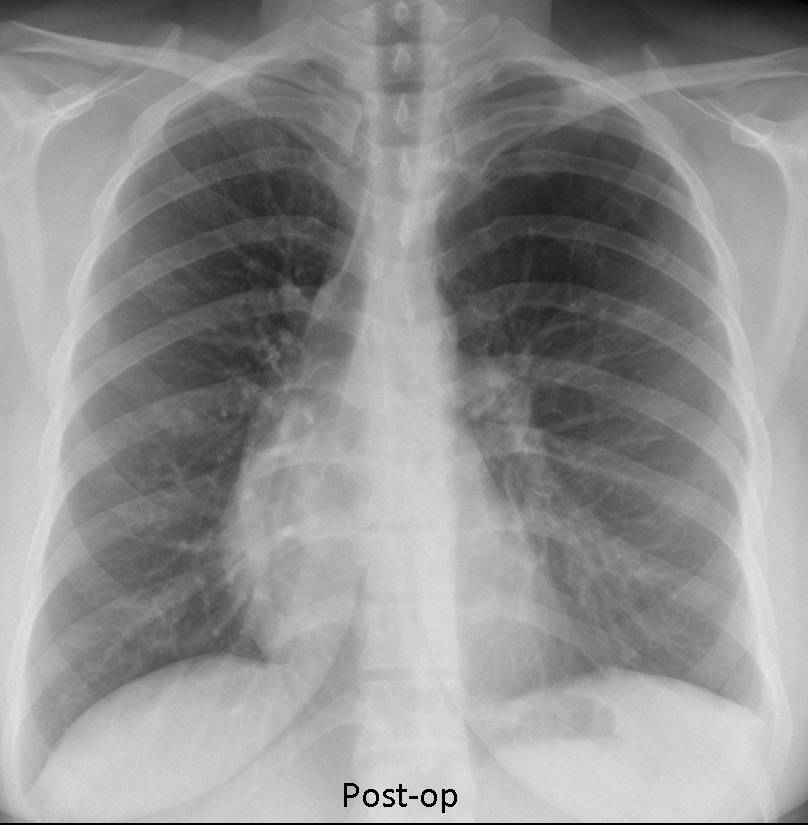
Fig. 3
I think most of you contributed with excellent discussions, but I will single out Dr. Sameh Khodair for his prompt and accurate answer.
Teaching point: this case represents an unusual cause of unilateral hyperlucent lung and complements case 47 of Dr. Pepe’s Diploma to be posted next Monday. Interesting to note that young patients with giant bullous emphysema are predisposed to lung cancer. I have personally seen three cases.