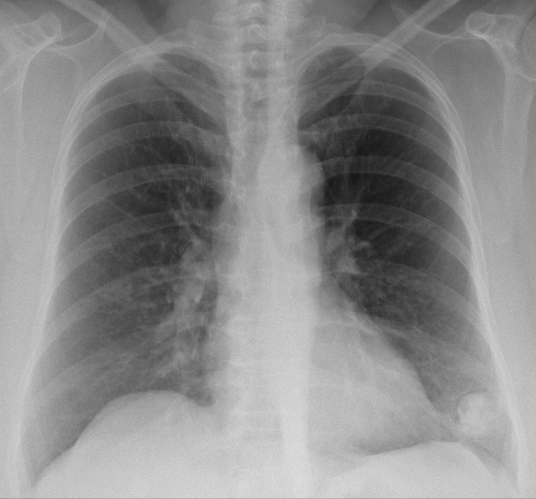
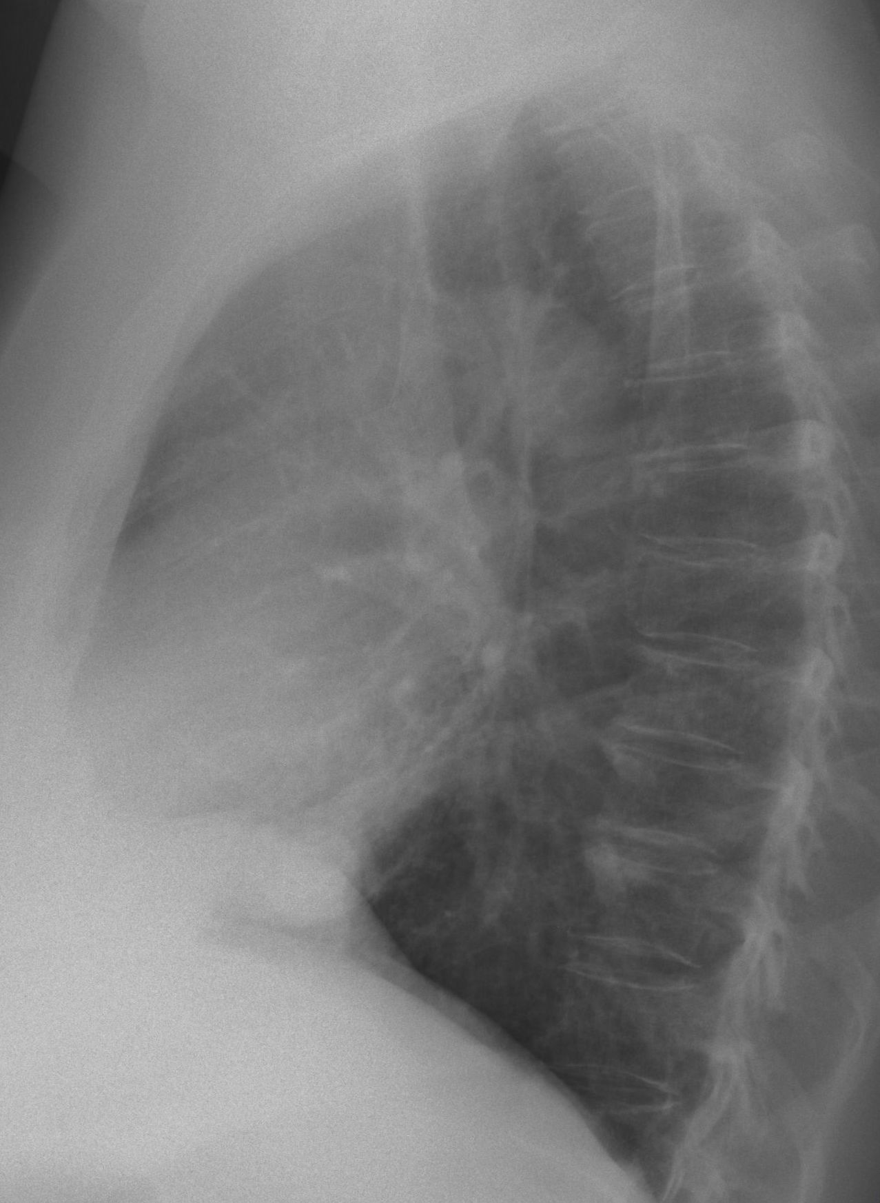
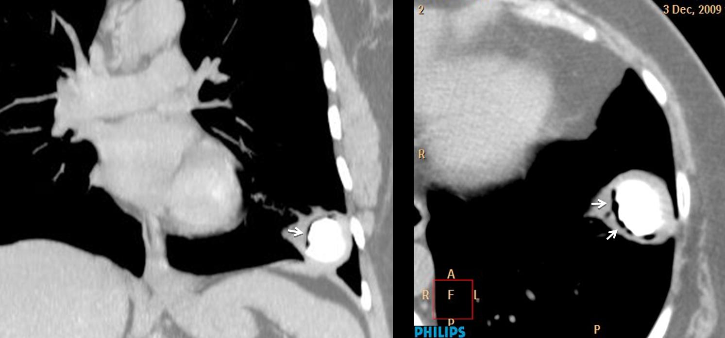
Muppet believes that the last week of the year deserves an unusual case. Images belong to a 61 year-old woman with frank haemoptysis for one week.
What would you say about the LLL lesion? Leave your thoughts and diagnosis in the comments section and come back on Friday for the answer.
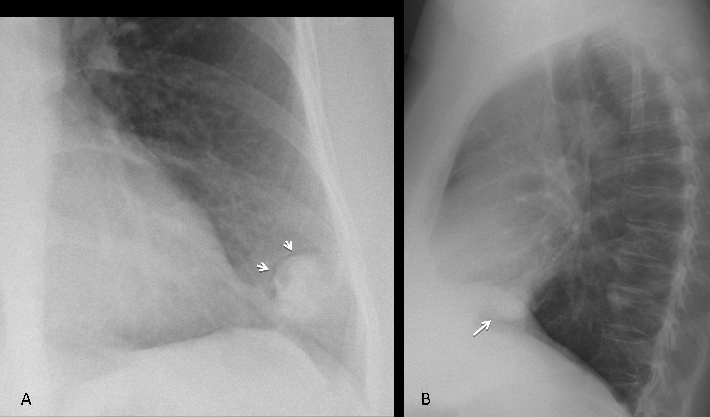
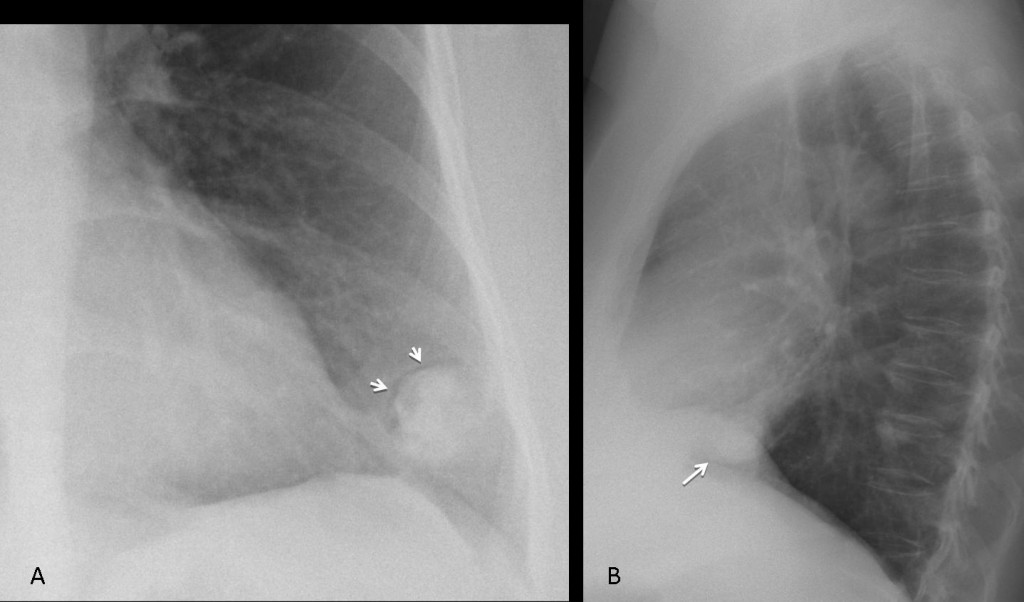
Findings: chest radiographs show a rounded lesion in the LLL with a visible crescent sign (A, arrows), indicating an occupied cavitary lesion. On close inspection, the cavitary content appears dense, suggesting that it may be calcified.
Coronal and axial CT confirms the air crescent (arrows) and a large calcification inside a cavity.
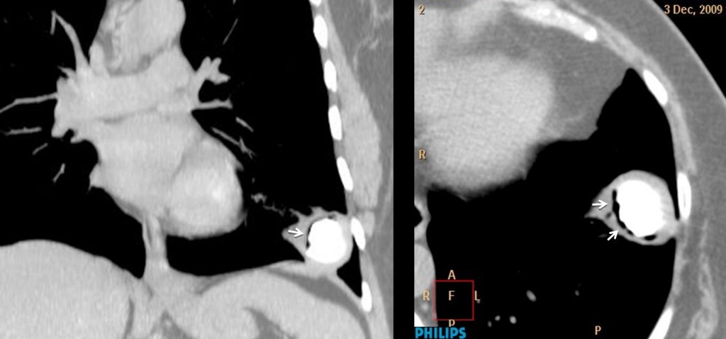
Final diagnosis: cavernolith
A cavernolith is defined as a calcified mass inside a lung cavity. Accepted etiology is a previous aspergillosis, which calcify with the passage of time. The pre-existing cavity is usually related to previous TB.
As you can imagine, this is my first and only cavernolith, which probably makes me an expert, since this is a rare finding. The interesting fact is that I saw this case four years ago, in 2009. The patient came to the ER with haemoptysis and the lesion was found. She refused surgery and was treated symptomatically. The haemoptysis disappeared and the patient has remained well since. A CT scan in 2013 did not show any change.
Congratulations to Lola la Piconera who made the diagnosis and to Genchi Bari, who pointed the way.
Teaching point: recognising the air crescent sign indicates a cavity with content and narrows the differential diagnosis. Perhaps in the future this sign will help you to diagnose your own cavernolith!