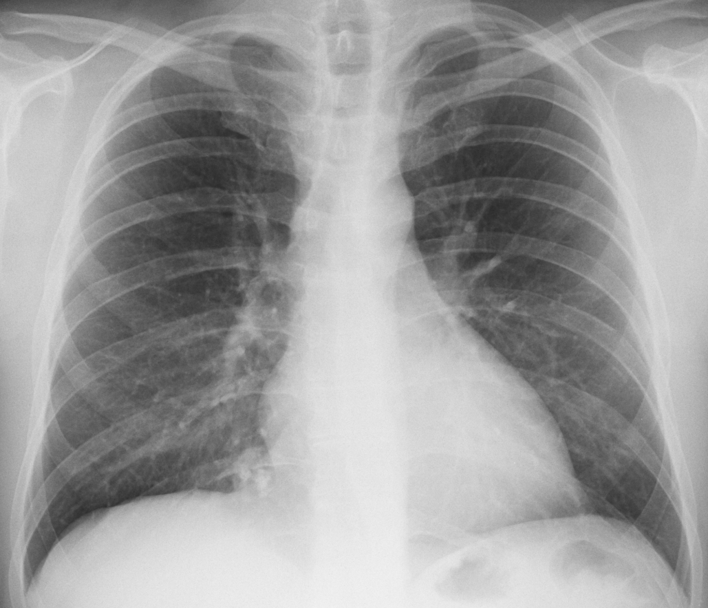
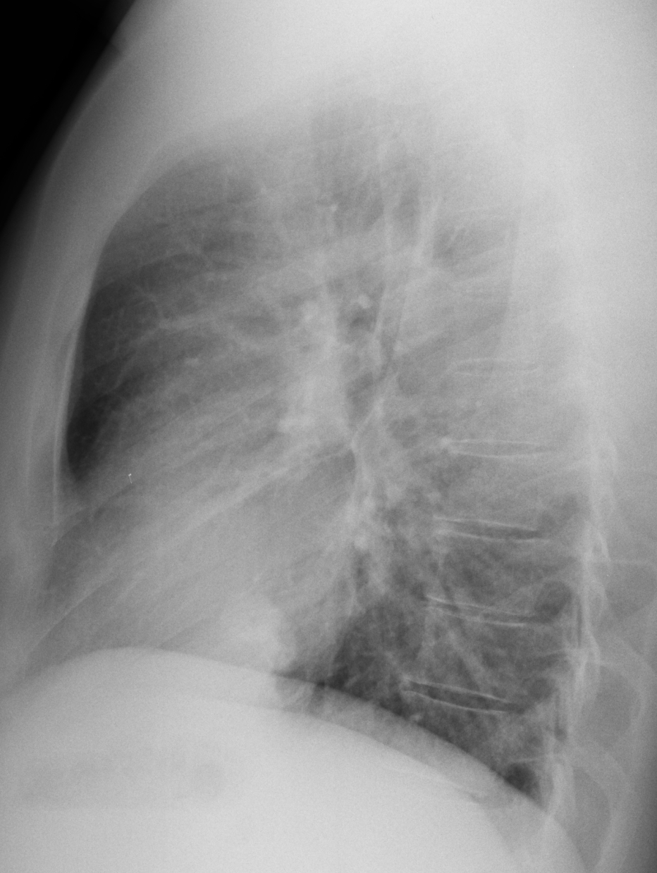
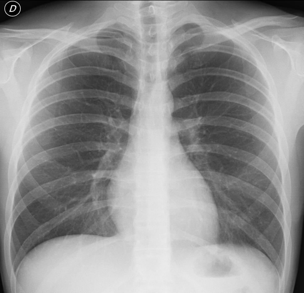
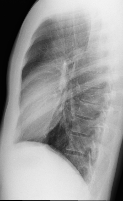
Since I am an old codger, I am only allowed to look at pre-op chest films. As one young member of staff told me: you may be better than us, but we are faster because we read all of them as normal!
Today I am showing two pre-op chests. Would you read them as normal? If not, what do you see? Leave your comments below and come back for the answer on Friday.
Patient 1 findings: there is a round pulmonary opacity at the right costophrenic angle (Fig 1 A,B arrow) with popcorn calcifications inside (A, red arrows).
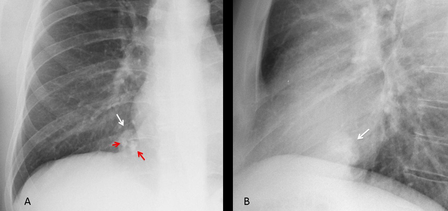
Fig. 1
The appearance is very suggestive of hamartoma, confirmed with axial and sagittal CT (Fig 2, arrows).
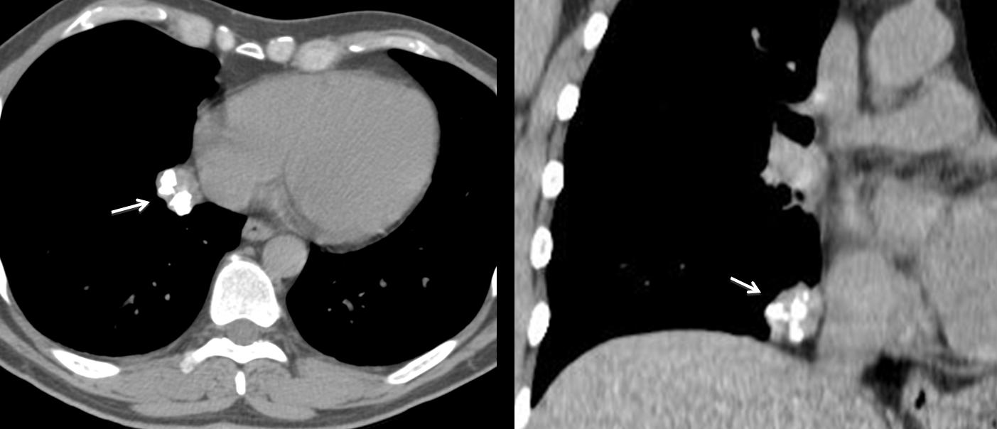
Fig. 2
Diagnosis: hamartoma of right lower lobe
Patient 2 findings: PA chest shows a ribbon-like structure (A, arrow) arising from the undersurface of the right tenth rib. It is barely visible in the lateral view, coursing anteriorly to the rib (B, arrow). The density of this structure is similar to that of the rib and, on close observation, cortical bone is visible.
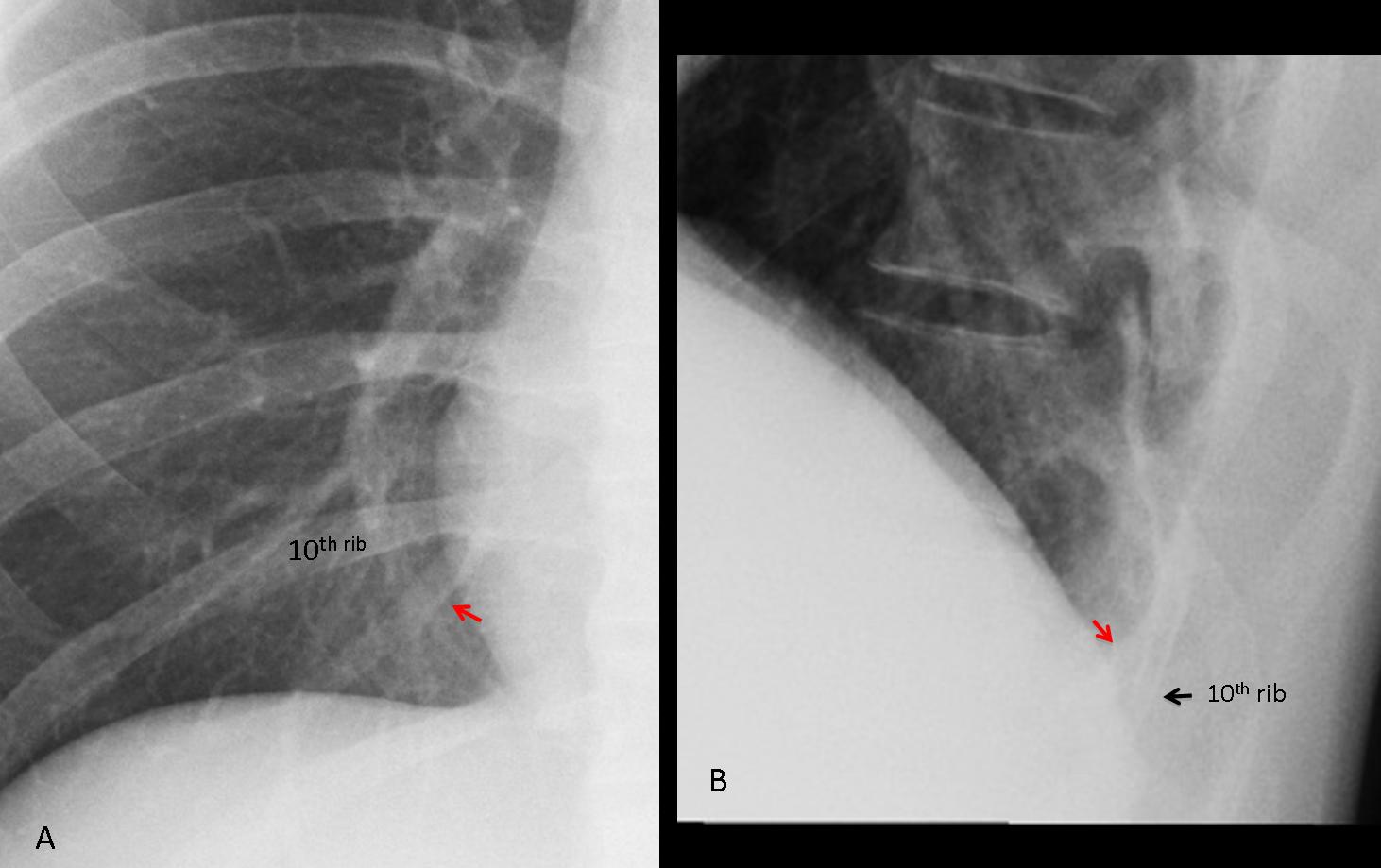
Patient 2
Diagnosis: intrathoracic rib. Because this entity has no clinical significance, no further studies were done.
Case 1 was reasonably easy to get. Case 2 was also easy if one recognises the density of the abnormality. As a matter of fact, several of you saw it, but confused it with an anomalous vein. Intrathoracic ribs are uncommon, more so if we fail to recognise them.
Teaching point: even apparently normal pre-op films may hide interesting findings. These two patients were seen last year.