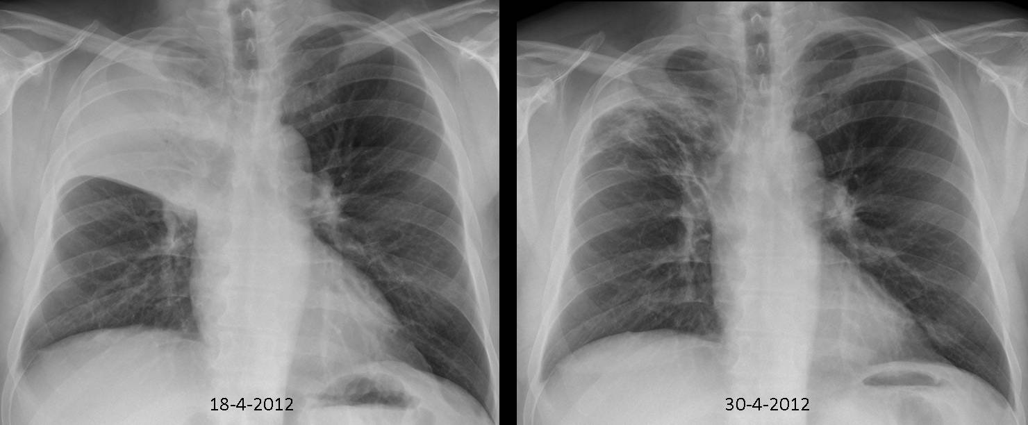
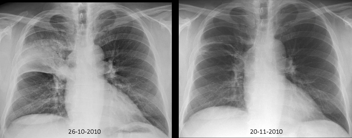
Dr. Pepe is still in Mexico, drinking tequila and enjoying mexican hospitality. He refuses to come back and asked me to cover for him. Radiographs belong to a 59-year-old male with fever. Had a similar episode two years ago which cleared with antibiotics.
1. Reactivation TB
2. Pneumonia
3. Carcinoma
4. None of the above
Findings: PA chest in 2012 shows an infiltrate that occupies the RUL with elevation of the minor fissure (Fig 1, arrow) indicating moderate collapse. Similar findings were present in 2010. After treatment, the infiltrates did not clear completely.
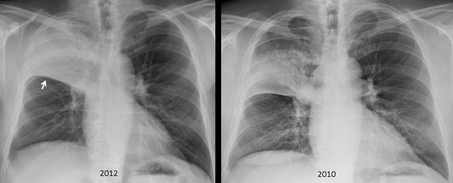
Fig. 1
The clue to the diagnosis lies in two facts: 1. Pneumonias that affect a whole lobe in adults (lobar pneumonias) should arouse suspicion of an endobronchial lesion; 2. Recurrent pneumonias in the same location should raise the possibility of an underlying condition, such as bronchiectasis, a congenital malformation or a lesion in the supplying bronchus. With this in mind, a CT should always be done. In this case, unenhanced CT shows an endobronchial lesion in the origin of the RUL bronchus (Fig 2, arrows). The time span (two years) goes against carcinoma. Surgery confirmed an endobronchial benign tumour.
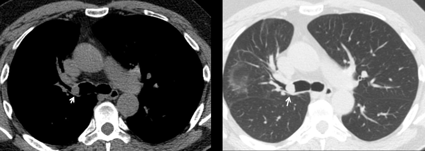
Fig. 2
Final diagnosis: Chondroma of RUL bronchus (possible chondromatous hamartoma)
Congratulations to Genchi Bari who offered an excellent discussion and suggested the diagnosis.
Teaching point: Lobar pneumonias in adults should clear completely after treatment. If not, an endobronchial lesion should be ruled out.