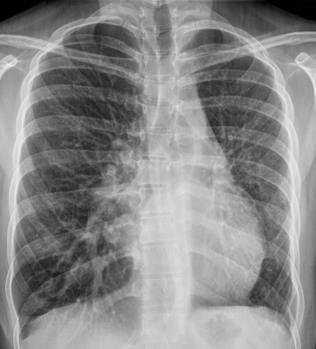
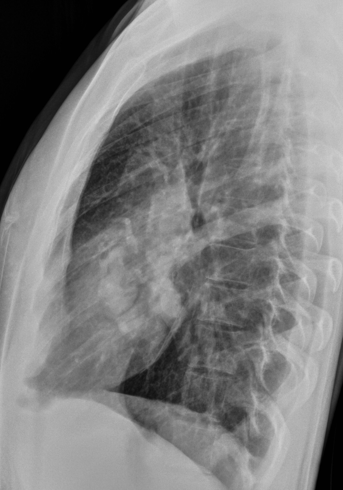
Dr. Pepe refuses to leave Mexico and I have to go there to bring him back. I have asked my friends Jose Vilar and Marisa Domingo to cover for me and present this week’s case. The radiographs below belong to an 18-year-old woman with recurrent haemoptysis.
Leave your thoughts and diagnosis in the comments section and come back for the answer on Friday.
1. Arteriovenous malformation
2. Hypoplasia of left lung
3. Vasculitis
4. None of the above
Figure 1 (PA and lateral radiographs):
a) the left lung has lost volume and there are few small vessels. There are no signs of air trapping or bronchiectasis. The right lung shows increased vascularity and a large vessel (
red arrow), b) in the lateral projection the left pulmonary artery is seen surrounding the left upper lobe bronchus (
blue arrows).
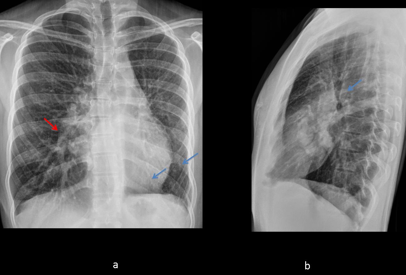
Fig. 1
Figure 2 (CT with contrast):
a) axial image shows a small left lung with thick septa (arrows) and absence of pulmonary veins, b) MIP coronal image shows large vessels in the right lung, small vessels and absence of pulmonary veins in the left.
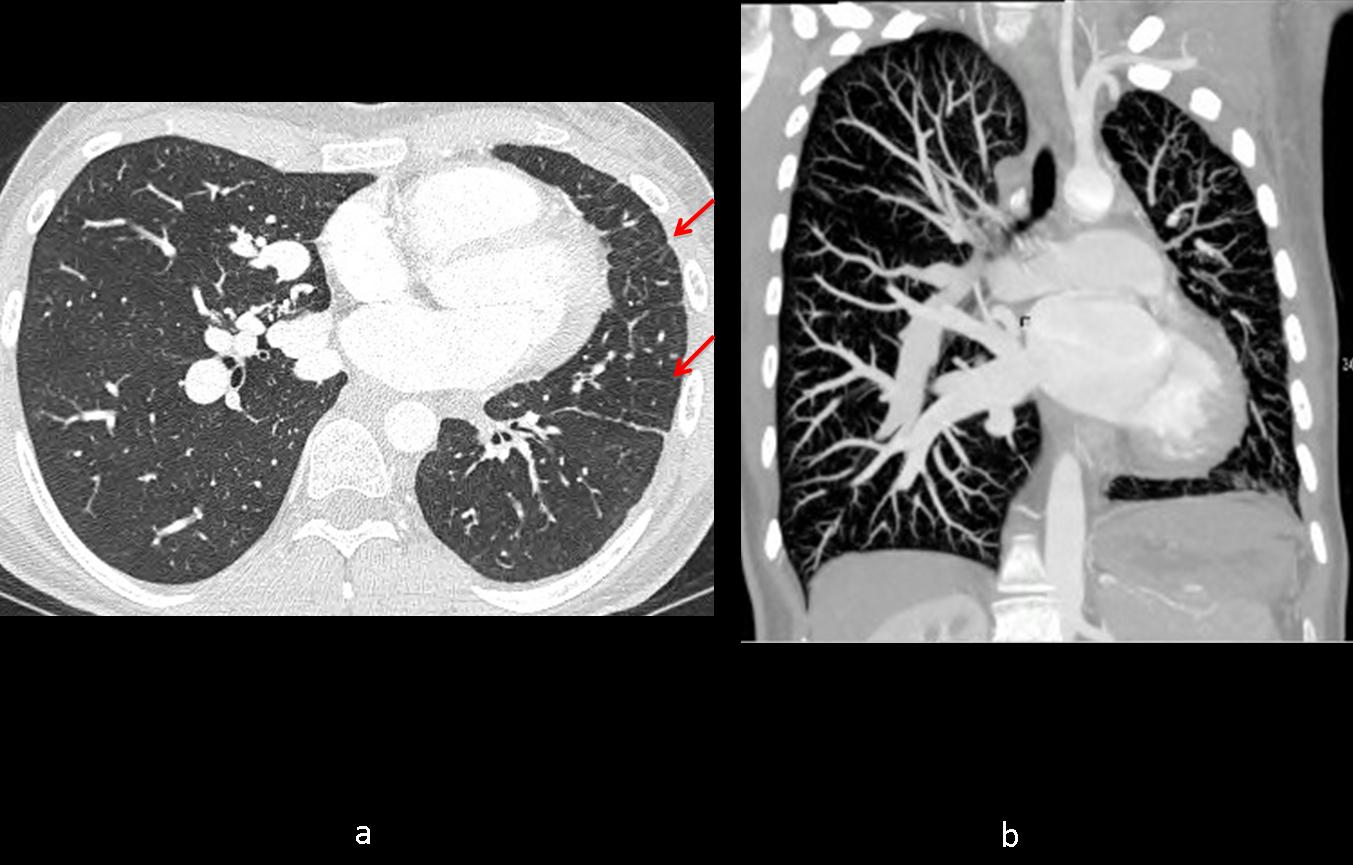
Fig. 2
Figure 3 (CT with contrast, reformated images):
a) coronal 3D image demonstrating absence of pulmonary veins in the left lung and a large unique vein in the right (white arrow), b) MIP coronal image shows tortuous (meandering) vein in the right lung (yellow arrow), absence of pulmonary veins in the left and enlarged bronchial arteries in the mediastinum (red arrows).
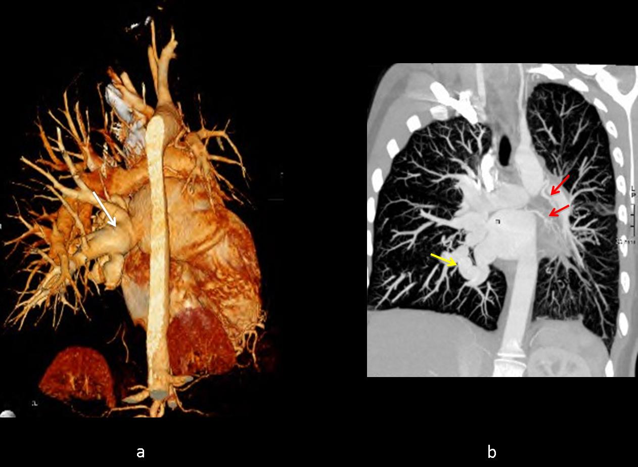
Fig. 3
Final diagnosis: congenital unilateral pulmonary vein atresia in the left lung and
large meandering vein in the right lung.
Teaching point: a small lung with a small pulmonary artery and septal lines is typical of congenital unilateral pulmonary vein atresia.