Dear Friends,
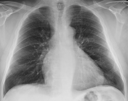
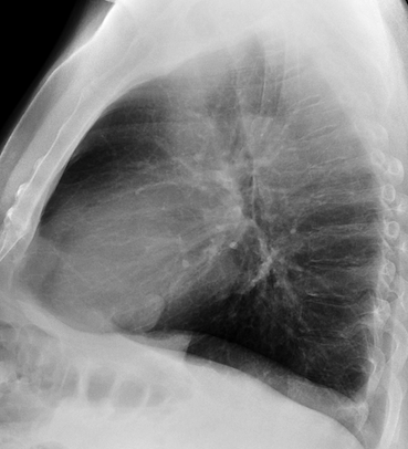
Our next case is that of a 60-year-old male with intermittent chest pain for the last year. If it is of any help, Muppet made the wrong diagnosis in this case. His mind was probably still in Mexico.
What do you see? What would be your diagnosis? Check the images below, leave your thoughts in the comments section, and come back on Friday for the answer.
Click here for the answer to case #87
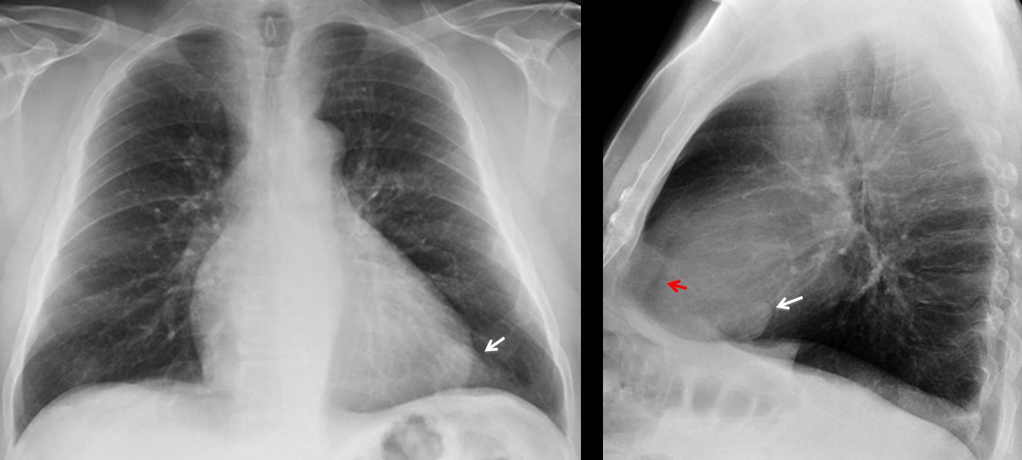
Findings: chest radiographs show a nodule adjacent to the cardiac silhouette (arrows). The epicardial fat is displaced posteriorly in the lateral view (red arrow) suggesting pericardial effusion.
With these findings I made a brilliant diagnosis: fibrous tumour of pleura irritating the adjacent pericardium and leading to pericardial effusion. CT proved me wrong, because the nodule was intrapulmonary (coronal CT, arrow). CT confirmed the pericardial effusion (sagittal CT, arrows) and the epicardial fat surrounded by it (red arrow).
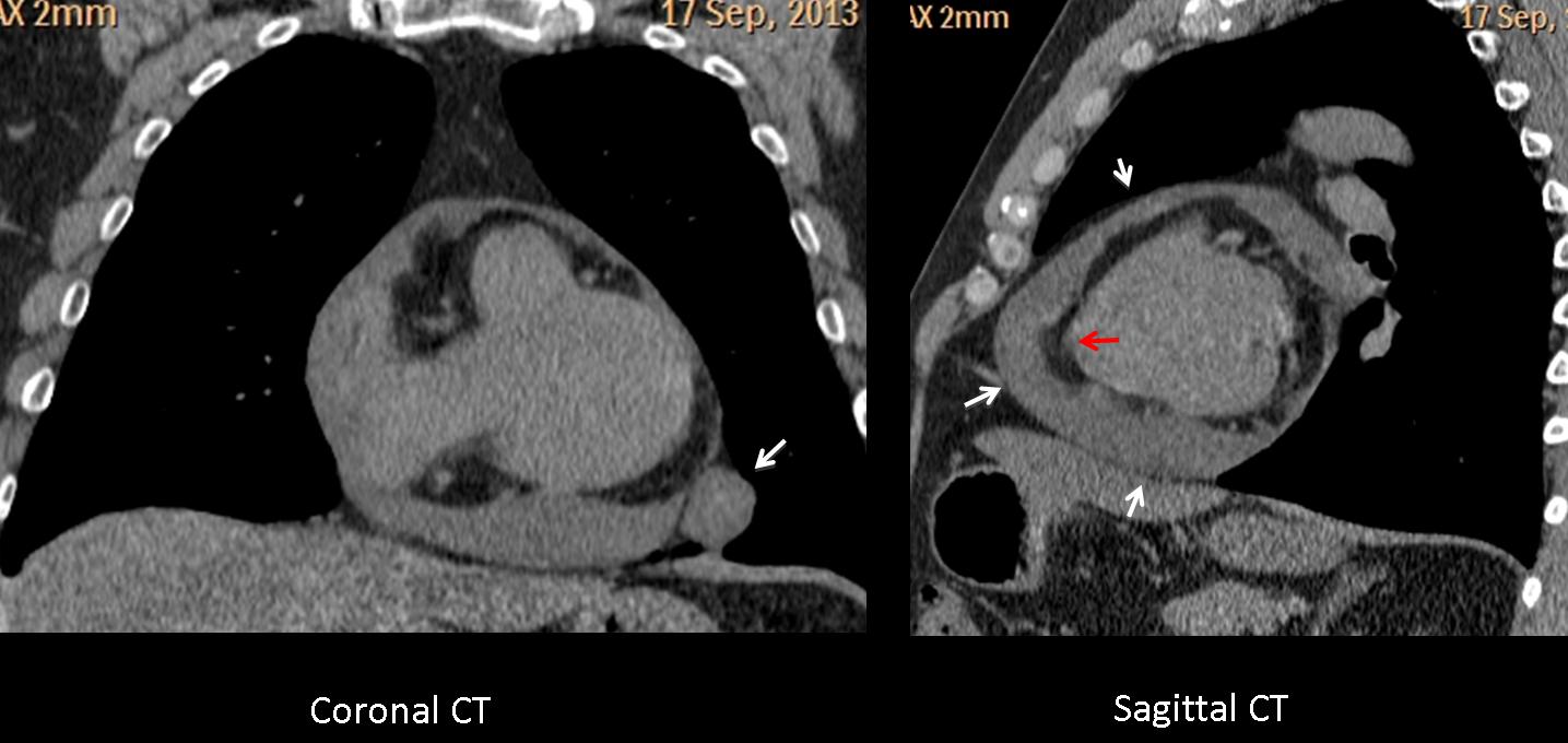
At surgery a benign hamartoma was found.
Final diagnosis: lung hamartoma with co-existing pericardial effusion.
Teaching point: This case is a good example of satisfaction of search: discovering the pericardial effusion raises the possibility of malignancy/ aggressive nodule. Pity I was wrong! To increase my misery, when I told one staff member that I had never seen a hamartoma with pericardial fluid, his answer was: “Now, you have”.


