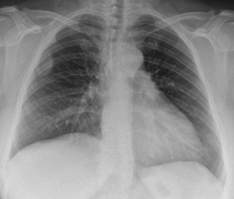
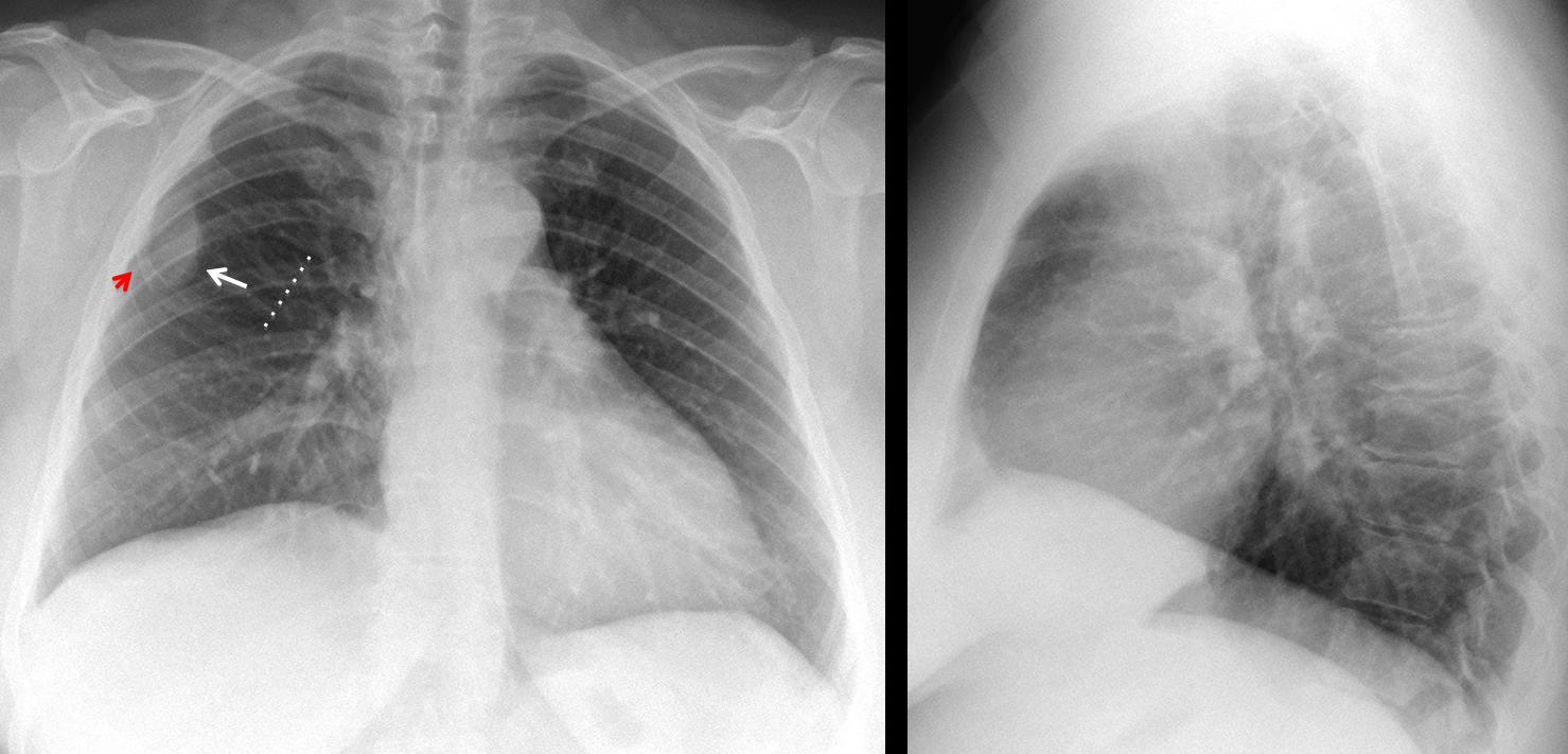
Muppet is concerned about the few responses in the last case. He wants to show an easy case to stimulate participation. Showing pre-op radiographs of a 41-year-old woman with rectal prolapse. The opacity in the right lung was not seen in the CT. What do you think?
Examine the images below, leave me your thoughts and diagnosis in the comments section, and come back on Friday for the answer.
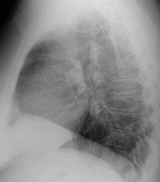
Findings: PA chest shows a right extrapulmonary opacity, accompanied by thinning of anterior right rib (red arrow). The intercostal space is widened (broken line). The lateral view is unremarkable.
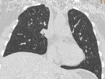
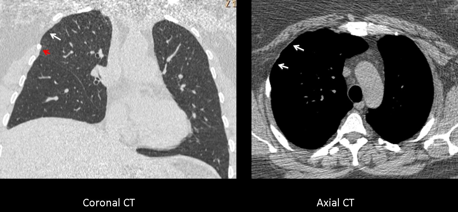
Coronal CT confirms the slender rib (red arrow) as well as the widened intercostal space (arrow) and atrophy of the intercostal muscles, confirmed with the axial CT (arrows).
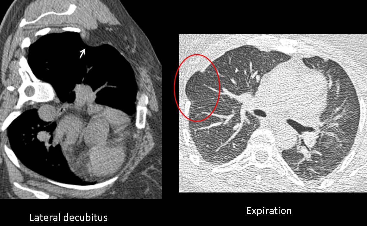
The striking finding is that the extrapulmonary opacity is not visible. Given the atrophic intercostal muscles, a good explanation is that the chest wall fat is herniating into the chest cavity. This theory was proved by placing the patient in the left decubitus position, which confirmed the fat herniation (arrow). An expiratory CT showed lung herniation in the same weak spot (red circle).
Interrogation of the patient discovered that she had surgery in childhood for complications of a pneumothorax.
Final diagnosis: herniation of chest wall fat into the thoracic cavity.
I want to congratulate Genchi Bari, Murzin and specially Gus, who made the definitive diagnosis
Teaching point: sometimes it is good to use our imagination to confirm suspected diagnosis. A little creativity can carry us a long way!