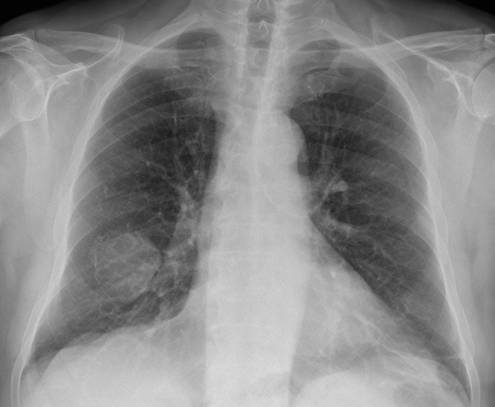
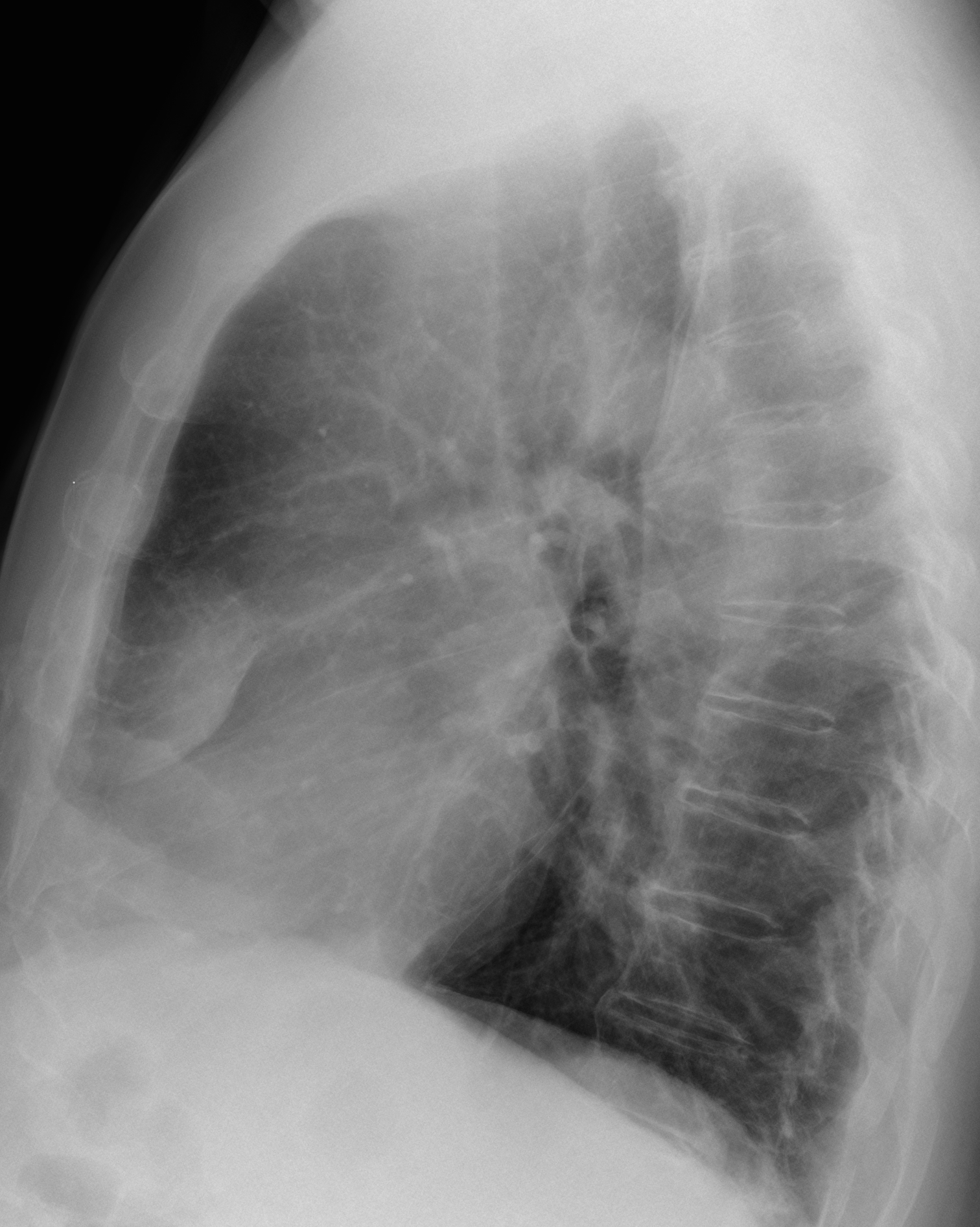
This week we are presenting pre-op radiographs of a 71-year-old man with torn ligaments of the right shoulder. As you can see, an unsuspected abnormality was found. What would be your diagnosis?
Check the images below, leave your thoughts in the comments section and come back on Friday for the answer.
1. Hydatid cyst
2. Carcinoma of the lung
3. Fibrous tumour of pleura
4. None of the above
Findings: there is a round opacity in the right lower lung, which appears to be extrapulmonary in the lateral view (B, arrow). Close observation of the PA chest discovers a finely calcified rim around the lesion (A, arrows).
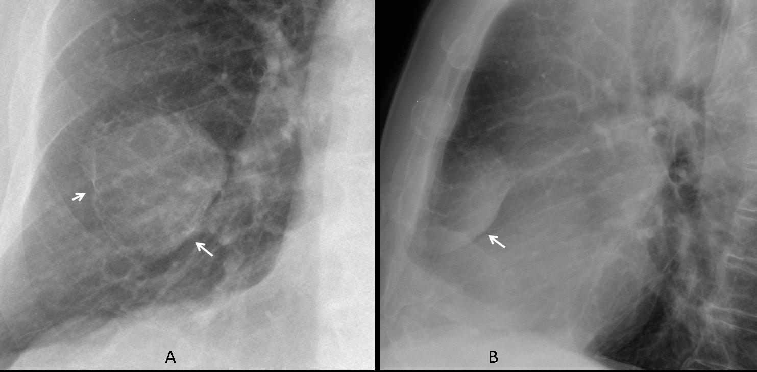
This finding excludes the three offered diagnoses and suggests a lesion arising from the anterior 5th rib which is poorly visualised compared to the one above and below.
Axial CT shows an expansive rib lesion with calcium inside (C, arrow) and a cortical break (D, arrow).
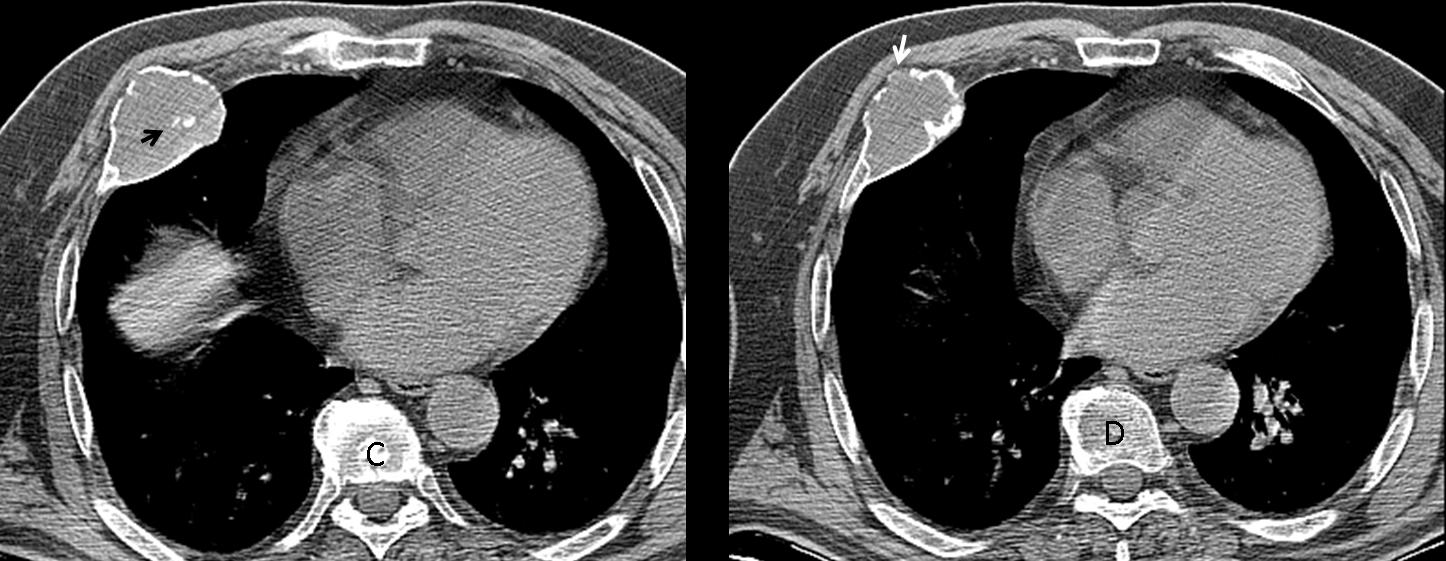
Final diagnosis: Grade II chondrosarcoma of the 5th rib.
Congratulations to Gus and Genchi Bari who, after some huffing and puffing, made the correct diagnosis.
Teaching point: When discovering an extrapulmonary lesion, always look at the underlying rib. In this particular case there was enough evidence to suspect that the lesion was originating in the rib.