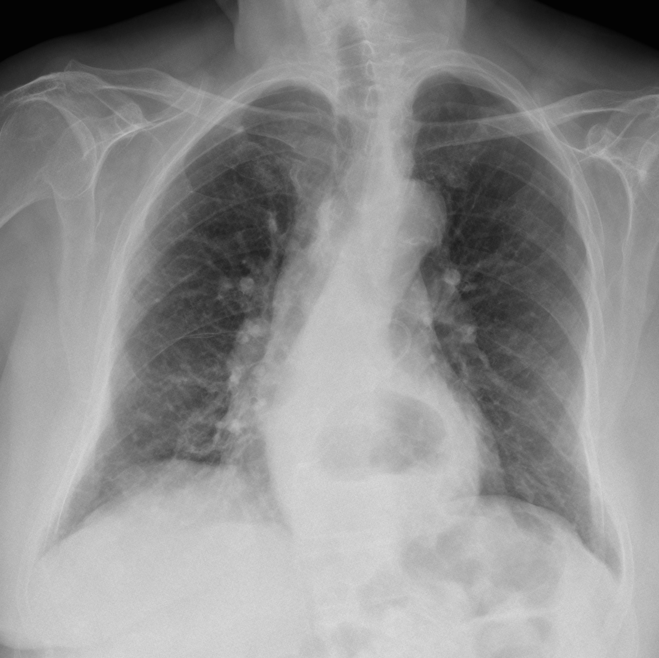
Presenting the case of a 85-year-old woman admitted with a severe urinary infection secondary to renal stones. Chest radiographs were taken one week after admission. Do you see any abnormalities in the chest?
Check the images below, leave your thoughts and diagnosis in the comments section and come back on Friday for the answer.
Findings: PA radiograph shows a pigtail catheter projected over the pulmonary artery silhouette (Fig 1A, arrow). The lateral view confirms that the catheter ends at the level of the main pulmonary artery (Fig 1B, red arrow) with its distal portion going into the abdomen (Fig 1B, arrows).
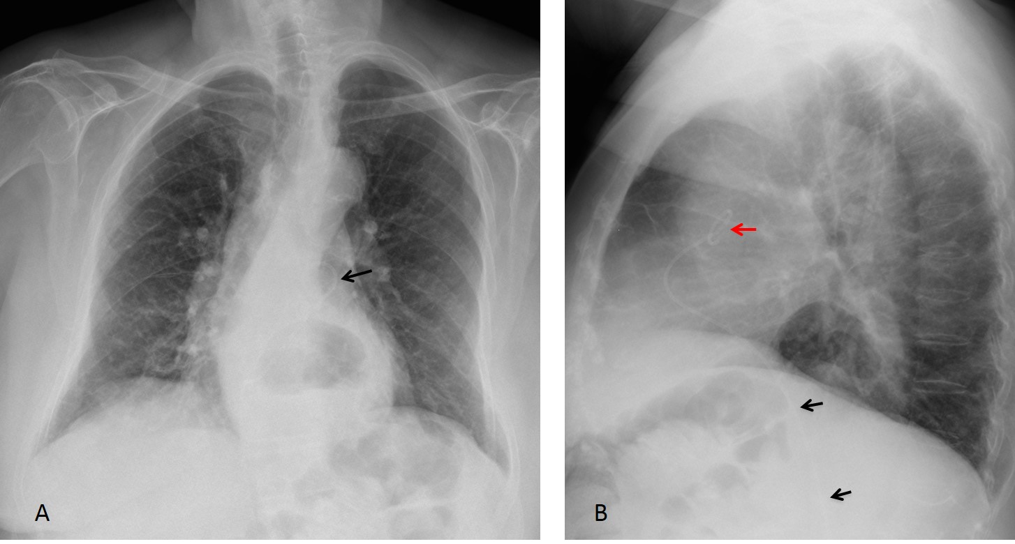
Fig. 1
Plain film of the abdomen shows the catheter in the midline (Fig 2, arrows).
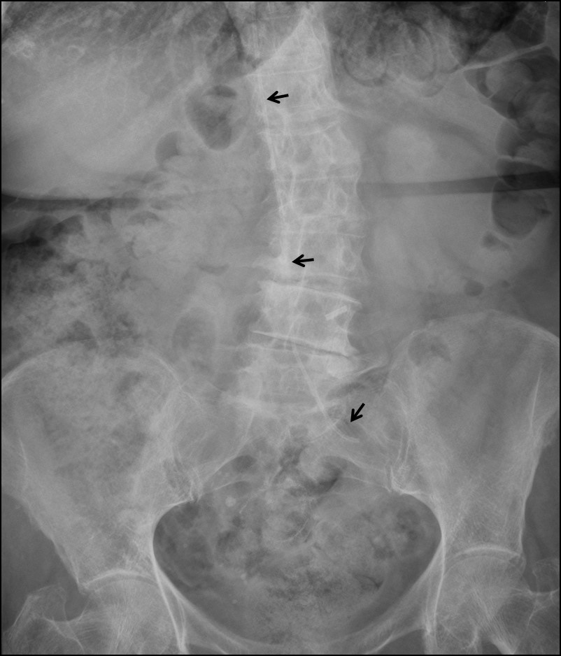
Fig. 2
Coronal and sagittal CT show the catheter entering the left iliac vein (Fig 3A, arrow), traversing the inferior vena cava (Fig 3A,B, arrows) and ending in the main pulmonary artery (Fig 3C, arrow).
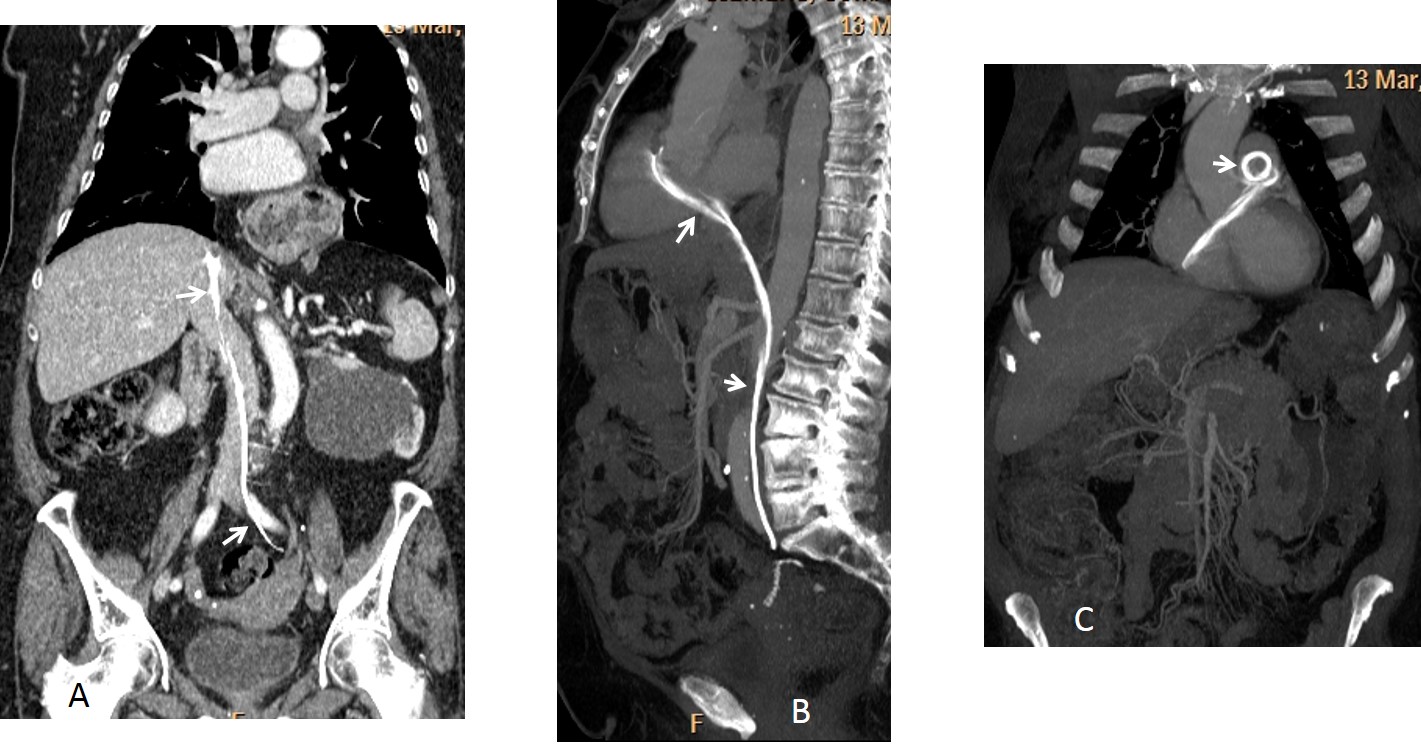
Fig. 3
In this patient, the pigtail was supposed to drain the left kidney. It was misplaced and perforated the left iliac vein, ending in the main pulmonary artery. Once discovered, it was removed percutaneously without complications.
Final diagnosis: inadvertent misplacement of urinary catheter into the inferior vena cava.
Congratulations to Jordi Velasquez, Genchi Bari and Francesco for their diagnostic acumen.
Teaching point: this is another case of catheter malposition. It is difficult to formulate a teaching point of this case, except for this: if anybody comes at you with a pigtail, cross your legs!