Dear Friends,
Dr. Pepe has urgent business (suspect he is visiting Miss Piggy) and asked me to cover for him this week and the next. He will be back with a new Diploma case on Monday, June 9.
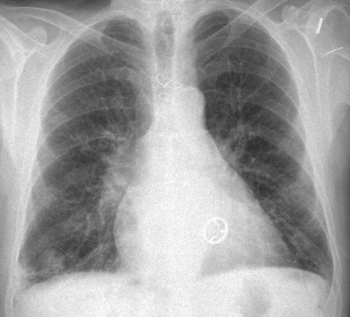
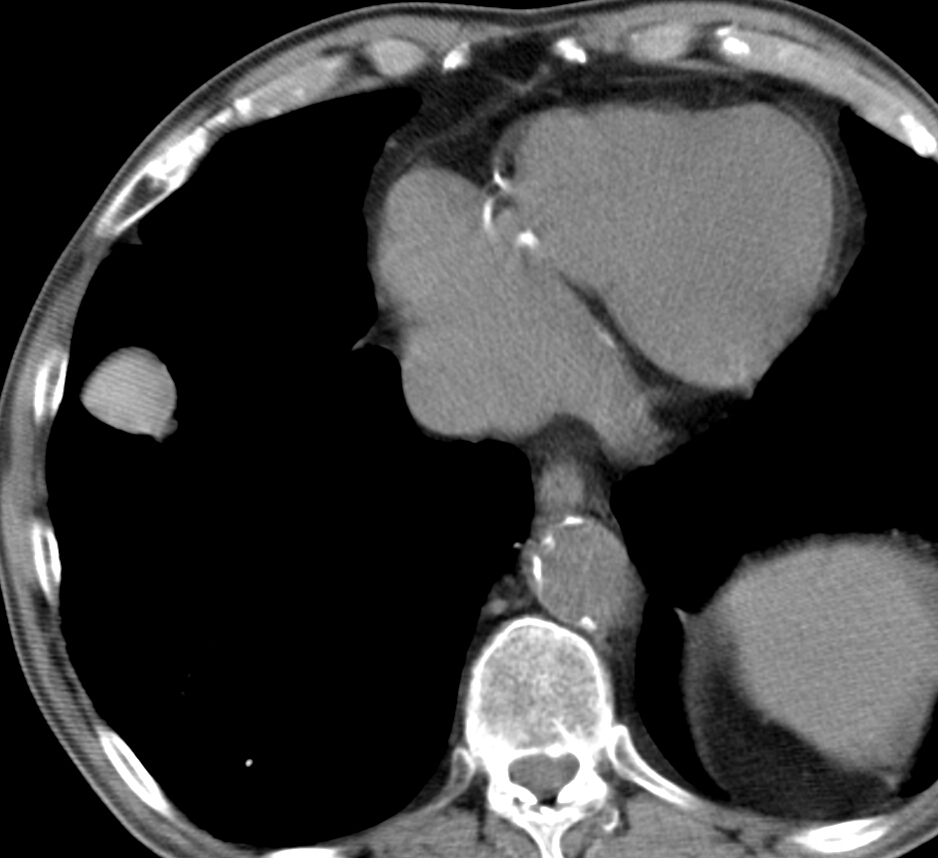
My good friend and former resident Dr. Eva Castañer has contributed with this week`s case. Images are of a 74-year-old male admitted with hemoptysis. Leave your diagnosis in the comments section and come back for the answer on Friday.
Diagnosis:
1. Pulmonary infarction
2. Carcinoma
3. Tuberculoma
4. None of the above
Click here for the answer to case #94
Findings: PA radiograph shows an ill-defined infiltrate at the right costophrenic angle and a nodular shadow at the right base (arrow). A prosthetic mitral valve is seen.
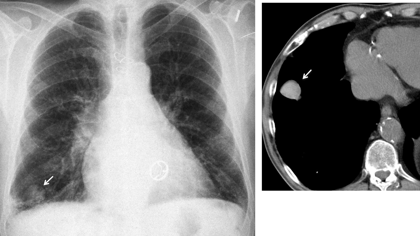
Unenhanced axial CT confirms a well-defined nodule (arrow), denser (84 HU) than the surrounding soft tissues. This finding raises the possibility of a haematoma, although a needle biopsy was done to exclude malignancy (negative result). Follow-up CT at one and four months shows a marked decrease in the size of the nodule.
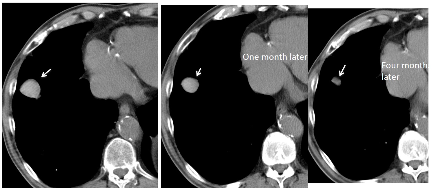
Final diagnosis: spontaneous lung haematoma in an anticoagulated patient.
Congratulations to Gaborini who was the very first to answer and gave the right diagnosis.
Teaching point: spontaneous haematomas are unusual but can be suspected by their high density at unenhanced CT. Follow-up films confirm the diagnosis by showing a steady decrease in size.


