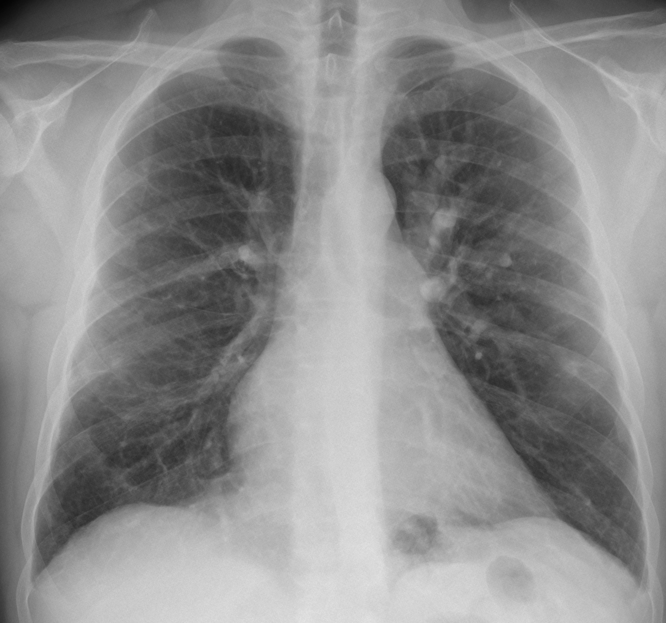
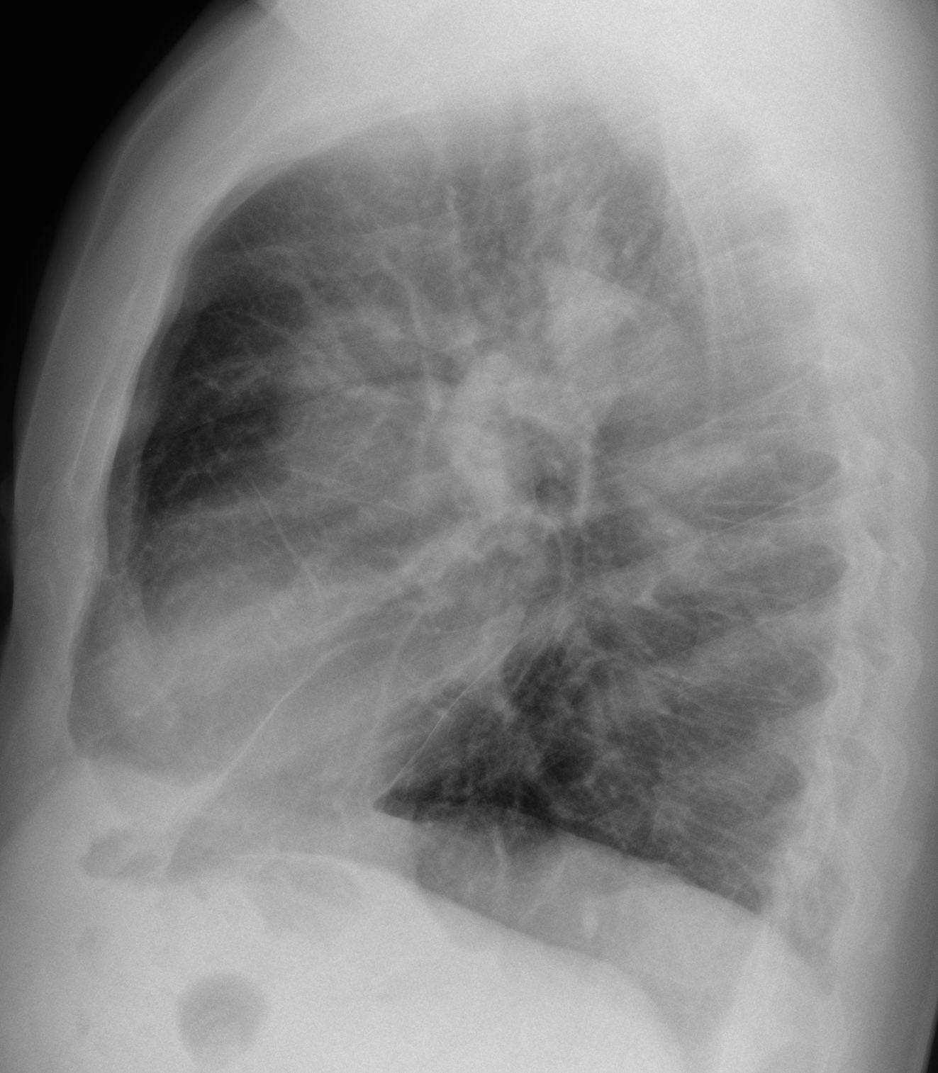
Muppet has insisted in presenting the following case: radiographs of a 48-year-old male with cognitive problems and episodes of amnesia. What do you see? What would be your diagnosis? Leave us your thoughts in the comments section and come back for the answer on Friday.
Findings: PA radiograph shows an obvious increase in size of the LUL vessels (Fig 1A, arrows). The lower lobes are oligemic and the calibre of vessels is decreased (Fig 1A, red arrows).
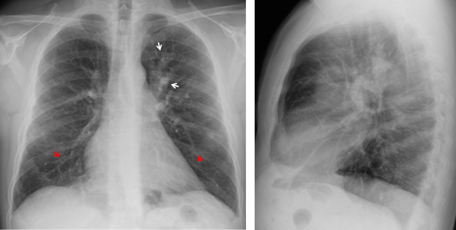
Fig. 1
These findings suggest a widespread vascular abnormality. The first possibility that comes to mind is chronic pulmonary embolism, but the hila are not enlarged and the heart looks normal. There are no signs of pulmonary emphysema, either. An uncommon possibility is pulmonary arteritis, supported by the clinical fact that the patient had neurologic symptoms. Further investigation discovered that the patient had been diagnosed with Takayasu’s disease eleven years earlier. At that time, CT (not available) discovered obliteration of lower lobe arteries and dilatation of LUL arteries. MRI was done and confirmed the findings (Fig 2).
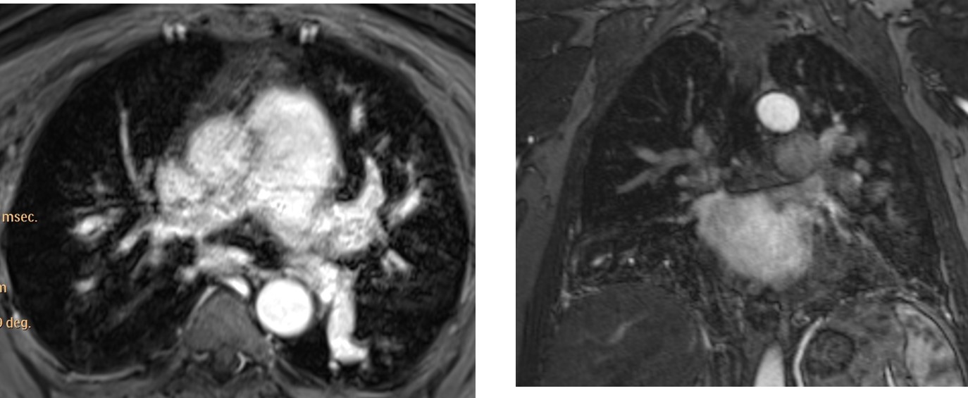
Fig. 2
Neck MRI showed occlusion of the left carotid artery (Fig 3, arrow).
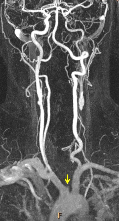
Fig. 3
Final diagnosis: Takayasu’s disease affecting pulmonary vessels.
Congratulations to Genchi Bari and Laurens who got close enough and deserve recognition.
I realise this is a difficult case, but Muppet made me do it! We have been waiting to show it ever since we saw the plain films in January. MRI was done two weeks ago and confirmed the findings.
Teaching point: vasculitis has variable manifestations. Think of vasculitis when several organs are affected and you don’t find a clear link.
This is the last case of the semester. I am leaving on vacation and Dr. Pepe will remain one more week to present the last Diploma case next Monday. I wish you all a happy summer vacation!