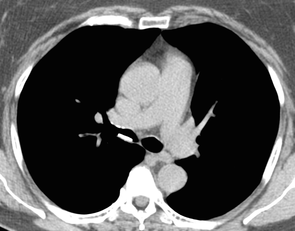
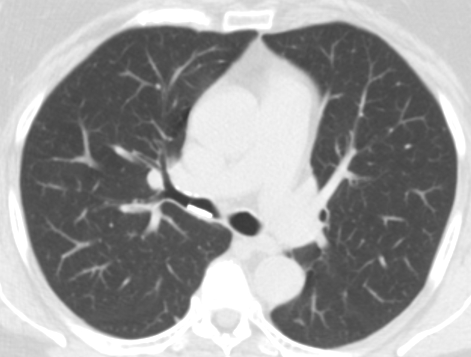
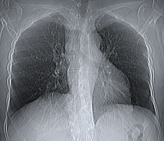
Muppet wants to start the new season with a warm-up case, provided by my good friend and former resident Carles Vilá. Images belong to a 62-year-old woman who has had a dry cough for the last six months. Chest radiograph was normal and a CT was taken.
1. Coin
2. Chicken bone
3. Pencil lead
4. None of the above
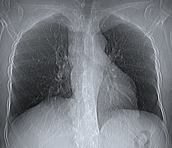
Findings: axial CT shows a linear metallic opacity within the intermediate bronchus (A, B arrows). Coronal CT reconstruction shows that the metallic opacity is rounded (C, arrow) and, in retrospect, can be seen in the scout film (D, arrows). At bronchoscopy a coin was found (insert).
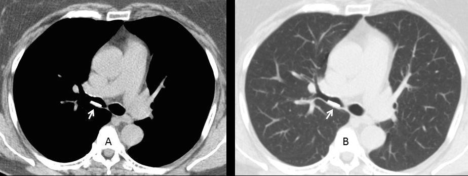
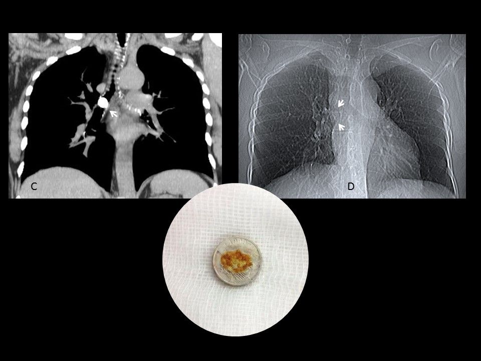
Final diagnosis: foreign body (coin) in intermediate bronchus.
Congratulations to Gus, who made an early and accurate diagnosis.
An interesting aspect of this case is the lack of detection of the coin in the chest radiograph and scout film. The coin is a Spanish peseta, made of aluminum with a weight of 0,55 grams and 14 mm of diameter. Its small size and aluminum’s low density makes the coin undetectable when imagined en face.
Another interesting point is that this coin was minted in 1999 and ceased circulating in 2001, after the arrival of the euro. Was it in the bronchus since 2001 until 2014 when it was discovered? To increase the mystery, the patient’s husband is a coin collector, casting doubts about the date of the aspiration.
Teaching point: remember that foreign bodies in adults are uncommon, but may happen, as proven by this case.