
Dear Friends,
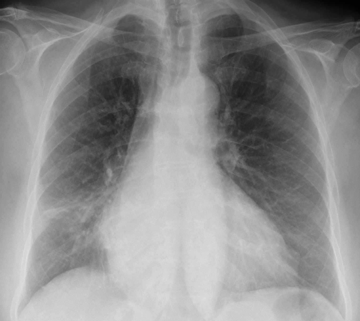
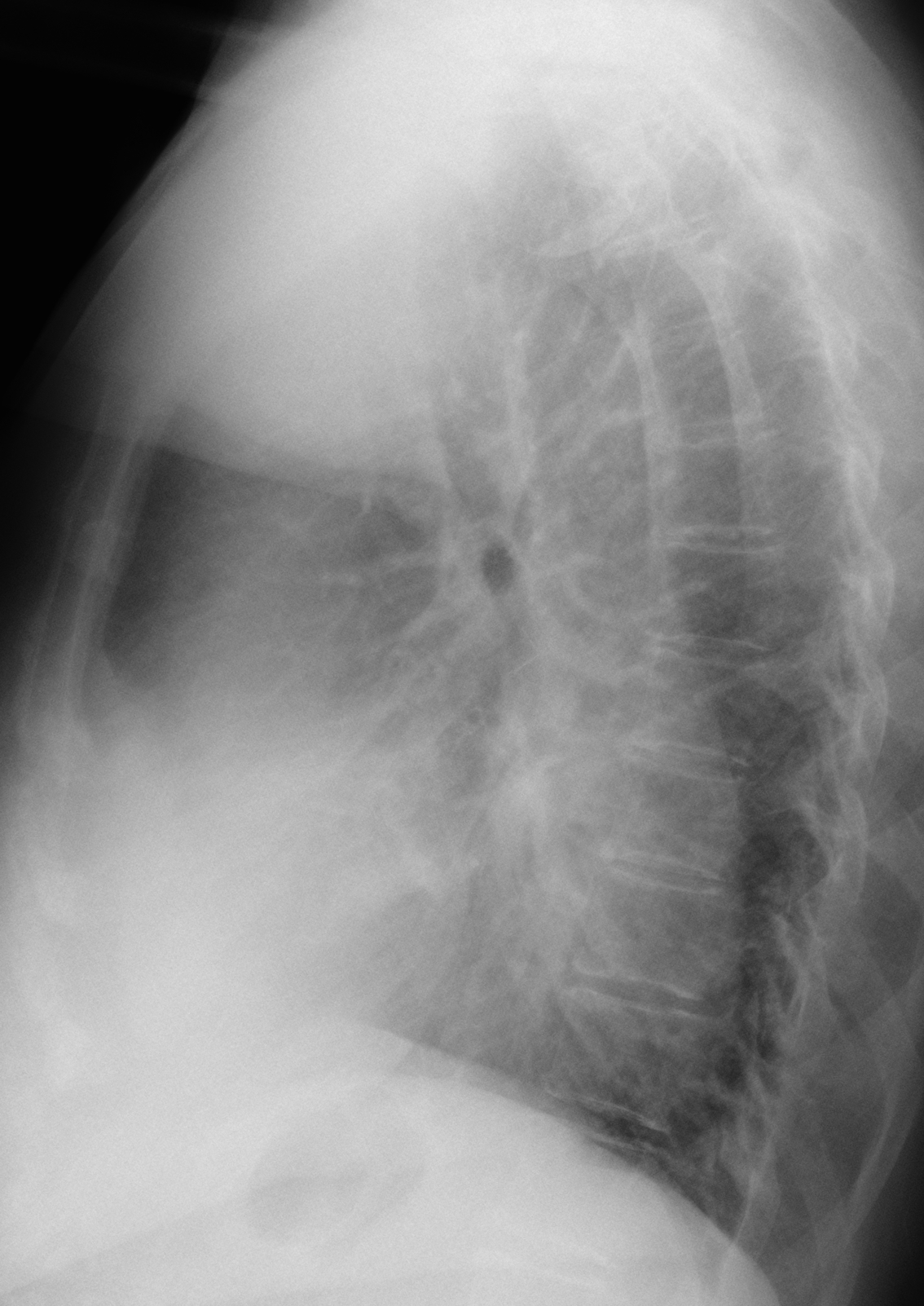
With your welfare in mind, Muppet has selected this week’s case. Radiographs belong to a 76-year-old woman, admitted to the hospital with moderate fever and vomiting.
Diagnosis:
1. Aspiration pneumonia
2. Tracheomegaly and pneumonia
3. Pneumonia and pericarditis
4. None of the above
Click here for the answer to case #98
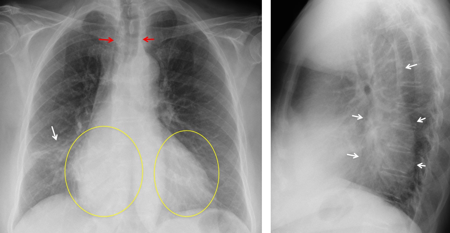
Findings: PA chest shows an RML infiltrate (arrow). There is also increased density of the right side of the heart compared to the left (circled areas), due to superposition of an elongated sausage-shaped density behind the heart in the lateral view (arrows). These findings are indicative of a dilated oesophagus, which is seen as an air-filled structure in the upper mediastinum (red arrows).
Coronal and sagittal CT confirms the dilated oesophagus (arrows), full of undigested food. Note the dilated air-filled upper oesophagus (red arrows) Oesophagoscopy demonstrated achalasia. No distal tumour was found.
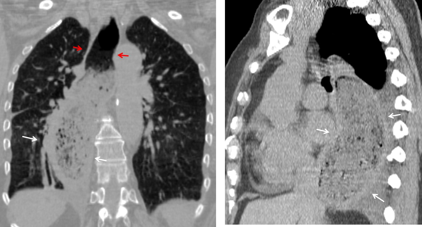
Final diagnosis: achalasia with aspiration pneumonia.
Congratulations to Adam and Jonathan who were the first to suggest the correct diagnosis.
Teaching point: when seeing a structure that traverses the middle mediastinum from top to bottom, always consider the oesophagus, with the descending aorta as the second possibility.


